Review
. 2017 Nov:71:3-12.
doi: 10.1016/j.semcdb.2017.06.018. Epub 2017 Jul 1.

How cells channel their stress: Interplay between Piezo1 and the cytoskeleton

Affiliations
Review

How cells channel their stress: Interplay between Piezo1 and the cytoskeleton

Jamison L Nourse et al. Semin Cell Dev Biol. 2017 Nov.

Abstract

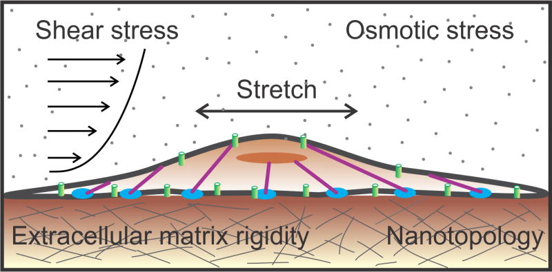
Cells constantly encounter mechanical stimuli in their environment, such as dynamic forces and mechanical features of the extracellular matrix. These mechanical cues are transduced into biochemical signals, and integrated with genetic and chemical signals to modulate diverse physiological processes. Cells also actively generate forces to internally transport cargo, to explore the physical properties of their environment and to spatially position themselves and other cells during development. Mechanical forces are therefore central to development, homeostasis, and repair. Several molecular and biophysical strategies are utilized by cells for detecting and generating mechanical forces. Here we discuss an important class of molecules involved in sensing and transducing mechanical forces - mechanically-activated ion channels. We focus primarily on the Piezo1 ion channel, and examine its relationship with the cellular cytoskeleton.

Keywords: Calcium signaling; Cytoskeleton; Mechanically-activated ion channels; Mechanotransduction; Piezo1; Traction forces.


Figures

Fig. 1
Fig. 1
Mechanical stimuli encountered by cells. Cells are subject to a variety of dynamic mechanical stimuli in the environment such as shear forces, osmotic stress, and stretch. They also sense mechanical cues in the matrix such as substrate rigidity and nanotopology (texture).
Fig. 2
Fig. 2
Piezo1 transduces outside-in mechanical forces. A variety of techniques have been developed to study transduction of outside-in mechanical forces by Piezo1. Some of these include: A) Membrane stretch elicited by suction pulses imparted by a high-speed pressure clamp in cell-attached patch clamp mode. B) Membrane stretch elicited by cell indentation with a glass probe controlled by a piezoelectric actuator in whole-cell patch clamp configuration. C) Shear stress induced by pulses of fluid flow from a perfusion pipette in whole-cell patch clamp configuration. D) Pulling or pushing on the cell surface by an AFM cantilever, while channel activity is measured by Ca2+ imaging on a confocal microscope. E) Cells are seeded on an array of microposts; a glass probe mounted on a piezoelectric actuator deflects a single micropost, mechanically stimulating a small number of channels in the vicinity of the micropost, while electrical activity is measured with whole-cell patch clamp. See Section 3 of the text for details on the techniques and results obtained. In all panels, actin filaments are shown in purple, focal adhesion zones in blue, Piezo1 molecules in green. Solid red arrows indicate force application; small broken arrows indicate ionic conduction through the channel.
Fig. 3
Fig. 3
Piezo1 transduces inside-out mechanical forces. A) Activation of Piezo1 by inside-out forces is studied by imaging Ca2+ influx through the channel with TIRFM. Since forces are generated by the cell itself, no external force stimulus is applied to the cell. B) Piezo1 is activated by traction forces (solid red arrow) generated at integrin-rich focal adhesion zones (blue) by myosin 2 molecules (black and yellow) along the actin cytoskeleton (purple). Broken red arrows denote ionic conduction through the channel. See Section 3 for details.

References

    1. Iskratsch T, Wolfenson H, Sheetz MP. Appreciating force and shape—the rise of mechanotransduction in cell biology. Nat. Rev. Mol. Cell Biol. 2014;15:825–833. - PMC - PubMed
    1. Coste B, Mathur J, Schmidt M, Earley TJ, Ranade S, Petrus MJ, Dubin AE, Patapoutian A. Piezo1 and Piezo2 are essential components of distinct mechanically activated cation channels. Science. 2010;330:55–60. - PMC - PubMed
    1. Yusko EC, Asbury CL. Force is a signal that cells cannot ignore. Mol. Biol. Cell. 2014;25:3717–3725. - PMC - PubMed
    1. Kung C, Martinac B, Sukharev S. Mechanosensitive channels in microbes. Annu. Rev. Microbiol. 2010;64:313–329. - PubMed
    1. Haswell ES, Phillips R, Rees DC. Mechanosensitive channels: what can they do and how do they do it? Structure. 2011;19:1356–1369. - PMC - PubMed

Publication types