. 2017 Aug 18;292(33):13541-13550.
doi: 10.1074/jbc.M116.772426. Epub 2017 Jul 4.

Kinetic and structural analyses reveal residues in phosphoinositide 3-kinase α that are critical for catalysis and substrate recognition

Affiliations

Kinetic and structural analyses reveal residues in phosphoinositide 3-kinase α that are critical for catalysis and substrate recognition

Sweta Maheshwari et al. J Biol Chem. .

Abstract

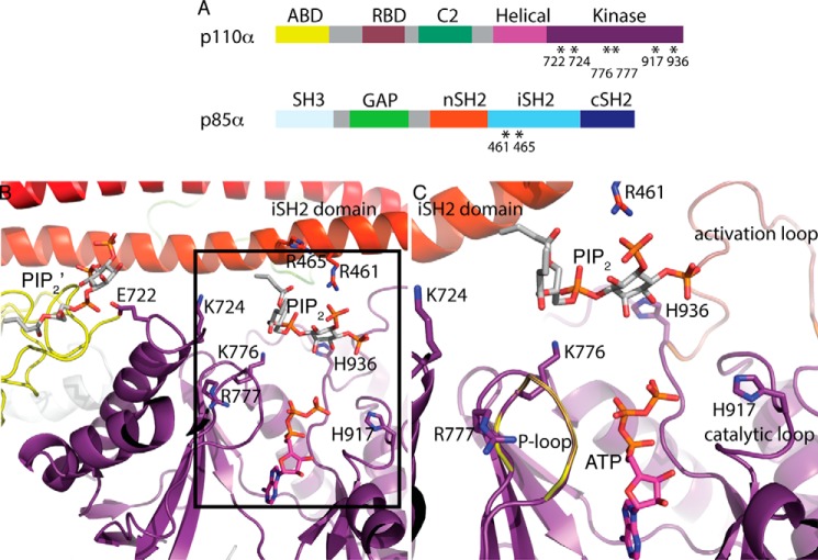
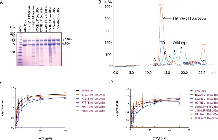
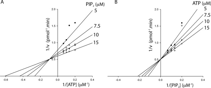
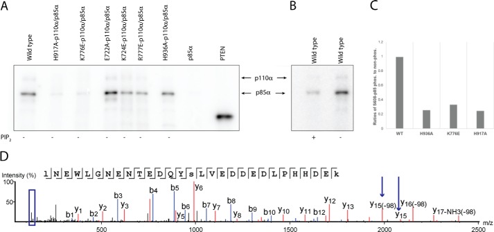
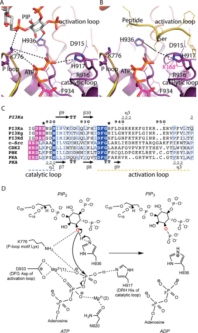
Phosphoinositide 3-kinases (PI3Ks) are ubiquitous lipid kinases that activate signaling cascades controlling cell survival, proliferation, protein synthesis, and vesicle trafficking. PI3Ks have dual kinase specificity: a lipid kinase activity that phosphorylates the 3'-hydroxyl of phosphoinositides and a protein-kinase activity that includes autophosphorylation. Despite the wealth of biochemical and structural information on PI3Kα, little is known about the identity and roles of individual active-site residues in catalysis. To close this gap, we explored the roles of residues of the catalytic domain and the regulatory subunit of human PI3Kα in lipid and protein phosphorylation. Using site-directed mutagenesis, kinetic assays, and quantitative mass spectrometry, we precisely mapped key residues involved in substrate recognition and catalysis by PI3Kα. Our results revealed that Lys-776, located in the P-loop of PI3Kα, is essential for the recognition of lipid and ATP substrates and also plays an important role in PI3Kα autophosphorylation. Replacement of the residues His-936 and His-917 in the activation and catalytic loops, respectively, with alanine dramatically changed PI3Kα kinetics. Although H936A inactivated the lipid kinase activity without affecting autophosphorylation, H917A abolished both the lipid and protein kinase activities of PI3Kα. On the basis of these kinetic and structural analyses, we propose possible mechanistic roles of these critical residues in PI3Kα catalysis.

Keywords: ATP; lipid kinase; p110a; p110ap85a; phosphatidylinositide 3-kinase (PI 3-kinase); phosphoinositide; protein kinase; serine/threonine protein kinase.


Conflict of interest statement

The authors declare that they have no conflicts of interest with the contents of this article

Figures

Figure 1.
Figure 1.
Structural basis of site-directed mutations in PI3Kα. A, domain organization of the p110α and p85α subunits of PI3Kα. RBD, Ras-binding domain; GAP, GTPase-activating protein. Mutated residues are indicated with asterisks. B, structure of the WT p110α/nip85α (Miller et al.; Ref. 31) in complex with the diC4-PIP2 lipid substrate (PDB ID 4OVV) and ATP modeled from the p110γ+ATP structure (PDB ID 1E8X). A second lipid-binding site (PIP2′) is shown. The kinase domain (with the activation, catalytic, and P-loops), the iSH2 domain and the ABD are colored in purple, red, and yellow, respectively. The amino acid residues mutated in this study (six in the p110α kinase domain and two in the p85α iSH2 domain) are illustrated as sticks. C, expanded view of the lipid substrate-binding site in the same orientation as B.
Figure 2.
Figure 2.
Lys-776, His-917, and His-936 on p110α are critical regulators of PI3Kα lipid kinase activity. A, SDS-PAGE analysis of PI3Kα constructs purified by anion-exchange chromatography. WT and eight mutants are shown. B, gel-exclusion chromatograms of the wild-type enzyme (shown in blue) and one of the mutants-H917A-p110α/p85α (shown in orange) as an example to illustrate the preservation of the oligomeric state of the mutants. Molecular weight standards (Bio-Rad) are shown in green, and peaks A, B, C, D, and E represent 678 kDa, 158 kDa, 44 kDa, 17 kDa, and 1.35 kDa, respectively. mAU, milliabsorbance units. C and D, comparison of lipid kinase activity of WT and mutants. Michaelis-Menten plots are shown with ATP (C) and PIP2 (D). Activity (V) is expressed in pmol/min. Error bars represent S.D.
Figure 3.
Figure 3.
Ternary complex formation is required for PI3Kα catalysis. A, Lineweaver-Burk plot of velocity versus ATP concentration while varying ATP concentrations (5–15 μm) at different fixed concentrations of PIP2 (5, 7.5, 10, and 15 μm). B, Lineweaver-Burk plot of velocity versus PIP2 concentration (5–15 μm) at different fixed concentrations of ATP (5, 7.5, 10, and 15 μm). The data were fitted to bisubstrate sequential models using SigmaPlot 13. Data shown are the mean of duplicate determinations from a single experiment and are representative of three such experiments.
Figure 4.
Figure 4.
Lys-776 and His-917 are required for autophosphorylation on p85α. A, SDS-PAGE analysis and autoradiography for WT and mutant PI3Kα proteins. Autophosphorylation of p85α using radiolabeled ATP as substrate in the absence of PIP2. The negative control is unphosphorylated p85α in the absence of p110α. The positive control is PTEN-phosphorylated by CK2 kinase. B, autophosphorylation of PI3Kα with and without PIP2. C, relative quantification of Ser-608 phosphorylation in p85α for WT PI3Kα and mutants by tandem mass spectrometry (MS/MS) of TMT-labeled peptides. D, tandem mass spectrum of TMT labeled peptides showing phosphorylation of Ser-608 (marked by arrows on y15) and reporter ions region (boxed in blue).
Figure 5.
Figure 5.
Structural insights into the catalytic mechanism of PI3Kα. Shown is the structure of the kinase domain of p110α harboring the P-loop and catalytic and activation loops with the lipid substrate (A) and the peptide substrate modeled from a cyclin-dependent protein kinase (CDK2) structure (B) (PDB ID 1QMZ). Lys-168 of PKA, shown in magenta, aligns with His-917 of p110α. C, sequence alignments of the catalytic loop (residues 912–920) and activation loop (residues 935–958) segments of class IA isoforms of PI3K (α, γ, δ) with different protein kinases (c-Src, tyrosine kinase; PHK, phosphorylase kinase; CDK2, cyclin-dependent kinase 2; PKA, protein kinase A). The conserved DFG motif of the activation loop and the XXDRH/HRDXK motif of the catalytic loop are highlighted in blue and purple, respectively. Mutated histidine residues His-917 and His-936 are marked with stars. D, molecular contacts between the substrates, conserved active-site residues, and the Mg2+ ions are shown. Asp-933 of the conserved DFG motif in the activation loop binds to the Mg2+ ion to orient the γ-phosphate of ATP for transfer. Asn-920 interacts with a second Mg2+ ion, which chelates the β-phosphate of ATP. Lys-776 of the conserved P-loop may support interactions with the γ-phosphate of ATP as well as 3′-OH of PIP2. His-936 of the activation loop is proposed to act as the catalytic base. His-917 of the conserved XXDRH motif in the catalytic loop, interacts with the γ-phosphate of ATP, and helps in the phosphoryl transfer step.

References

    1. Fruman D. A., Meyers R. E., and Cantley L. C. (1998) Phosphoinositide kinases. Annu. Rev. Biochem. 67, 481–507 - PubMed
    1. Chang H. W., Aoki M., Fruman D., Auger K. R., Bellacosa A., Tsichlis P. N., Cantley L. C., Roberts T. M., and Vogt P. K. (1997) Transformation of chicken cells by the gene encoding the catalytic subunit of PI 3-kinase. Science 276, 1848–1850 - PubMed
    1. Cho H., Mu J., Kim J. K., Thorvaldsen J. L., Chu Q., Crenshaw E. B. 3rd, Kaestner K. H., Bartolomei M. S., Shulman G. I., and Birnbaum M. J. (2001) Insulin resistance and a diabetes mellitus-like syndrome in mice lacking the protein kinase Akt2 (PKBβ). Science 292, 1728–1731 - PubMed
    1. Maira S. M., Finan P., and Garcia-Echeverria C. (2010) From the bench to the bed side: PI3K pathway inhibitors in clinical development. Curr. Top. Microbiol. Immunol. 347, 209–239 - PubMed
    1. Koorella C., Nair J. R., Murray M. E., Carlson L. M., Watkins S. K., and Lee K. P. (2014) Novel regulation of CD80/CD86-induced phosphatidylinositol 3-kinase signaling by NOTCH1 protein in interleukin-6 and indoleamine 2,3-dioxygenase production by dendritic cells. J. Biol. Chem. 289, 7747–7762 - PMC - PubMed

MeSH terms