. 2017 Sep;24(9):459-474.
doi: 10.1530/ERC-16-0340. Epub 2017 Jul 4.

Functional consequences of the first reported mutations of the proto-oncogene PTTG1IP/PBF

Affiliations

Functional consequences of the first reported mutations of the proto-oncogene PTTG1IP/PBF

W Imruetaicharoenchoke et al. Endocr Relat Cancer. 2017 Sep.

Abstract

Pituitary tumor-transforming gene 1-binding factor (PTTG1IP; PBF) is a multifunctional glycoprotein, which is overexpressed in a wide range of tumours, and significantly associated with poorer oncological outcomes, such as early tumour recurrence, distant metastasis, extramural vascular invasion and decreased disease-specific survival. PBF transforms NIH 3T3 fibroblasts and induces tumours in nude mice, while mice harbouring transgenic thyroidal PBF expression show hyperplasia and macrofollicular lesions. Our assumption that PBF becomes an oncogene purely through increased expression has been challenged by the recent report of mutations in PBF within the Catalogue of Somatic Mutations in Cancer (COSMIC) database. We therefore sought to determine whether the first 10 PBF missense substitutions in human cancer might be oncogenic. Anisomycin half-life studies revealed that most mutations were associated with reduced protein stability compared to wild-type (WT) PBF. Proliferation assays narrowed our interest to two mutational events which significantly altered cell turnover: C51R and R140W. C51R was mainly confined to the endoplasmic reticulum while R140W was apparent in the Golgi apparatus. Both C51R and R140W lost the capacity to induce cellular migration and significantly reduced cell invasion. Colony formation and soft agar assays demonstrated that, in contrast to WT PBF, both mutants were unable to elicit significant colony formation or anchorage-independent growth. However, C51R and R140W retained the ability to repress radioiodide uptake, a functional hallmark of PBF. Our data reveal new insight into PBF function and confirm that, rather than being oncogenic, mutations in PBF are likely to be passenger effects, with overexpression of PBF the more important aetiological event in human cancer.

Keywords: COSMIC; PTTG1IP; TCGA; mutation; proliferation.


Figures

Figure 1
Figure 1
The genomic landscape of PBF in cancer. (A) Alteration frequency (>0%) of significant events in PBF, such as non-synonymous mutations, high-level amplification and homozygous deletion, in multiple human cancers in the TCGA database. (B) Alteration frequency of all events in PBF, including heterozygous loss and allelic gain, across all human cancer datasets with Copy Number Alteration information in the TCGA database. Inset – the distribution of all types of alteration to PBF. (C) Schematic diagram of PBF highlighting the first ten PBF mutations reported in the COSMIC database and their localisation within its functional domains (PSI, plexin–semaphorin–integrin; NLS, nuclear localisation signal). Below: amino acid conservation of the mutated residues across 6 animal species. *, identical/fully conserved amino acid; :, similar/conserved residues according to Clustal Omega analysis (http://www.ebi.ac.uk/Tools/msa/clustalo/).
Figure 2
Figure 2
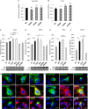
Expression, localisation and half-life of PBF mutations. (A) Subcellular localisation of each HA-tagged mutant (green) within HeLa cells using an anti-HA antibody. Blue indicates nuclear DAPI staining. As HA-tagged R140W is difficult to detect through immunofluorescent microscopy, FLAG-tagged wild-type (WT) and R140W PBF were stained using an anti-FLAG antibody. Magnification = 100×. Bars = 20 µm. (B) Anisomycin half-life assays in MCF7 cells transfected with HA-tagged WT PBF and 9 mutations and treated for 0, 12 or 24 h, with quantification of relative PBF levels shown in Western blotting (FLAG-tagged WT and R140W are shown alongside). (C) Quantification of protein stability of the ~25–37 kDa isoforms of PBF in MCF7 cells following normalisation to β-actin expression. Data presented as mean values ± s.e. (n = 2). (D) SIFT scores of all ten PBF mutations, where scores <0.05 are predicted to represent deleterious amino acid substitutions, which are defined as ‘damaging.’ (E) Western analysis of lysate from COS-7 cells transfected with vector only (VO), WT PBF and the 10 PBF mutants showing the expression levels and multiple forms of the HA-tagged PBF proteins. (F) PNGase F treatment of lysate from COS-7 cells transfected with WT PBF-HA results in a band at ~20 kDa and confirms N-linked glycosylation of PBF. PBF with a double substitution of N45 and N54 for alanine is also detected at ~20 kDa and cannot be modified by PNGase F confirming these are the only sites of glycosylation. (G) Western blotting of lysate from COS-7 cells transfected with HA-tagged PBF with discrete mutation of N45 and N54 suggests that both sites are glycosylated. (H) Western analysis of WT PBF-HA expressed in HeLa cells using non-reducing (minus β-mercaptoethanol (β-ME)) conditions clearly reveals the presence of a dimer ~50 kDa. (I) Western blotting demonstrating the presence of the ~50 kDa band, and therefore the ability of FLAG-PBF to form dimers, in COS-7, HeLa and MCF7 cells transfected with WT FLAG-PBF and VO control. (J) Proximity ligation assays (PLAs) demonstrating an interaction between FLAG-PBF and PBF-HA in HeLa cells (red spots indicate <30–40 nm distance between the anti-FLAG and anti-HA antibody epitopes and signify protein–protein interaction). VO co-transfections with either FLAG-PBF or PBF-HA represent negative controls.
Figure 3
Figure 3
The influence of PBF mutations on thyroid and breast cell proliferation. MTS proliferation assays in SW1736 (A) and TPC1 (B) thyroid cancer cells demonstrated that 3 substitution mutations at residue G106 failed to significantly alter mitochondrial activity, compared with WT PBF, as a marker of cell number. BrdU cellular proliferation experiments in TPC1 (C) and MCF7 (D) cells transfected with VO, WT PBF, C51R, V55I, R140W and R146W mutants. MTS proliferation assays in TPC1 (E) and MCF7 (F) cells transfected with VO, WT PBF, C51R and R140W substitutions. Beneath – Western blots probed with anti-PBF antibody and β-actin as loading control demonstrate PBF expression to assess transfection. Data presented as mean values ± s.e. (n = 3). *P < 0.05, **P < 0.01, ***P < 0.001. Representative immunofluorescent microscopy of FLAG-tagged PBF and C51R and R140W mutants (green) and ER marker (anti-PDI Ab, red) or Golgi marker (anti-golgin-97 Ab, red) with subcellular co-localisation (yellow) in HeLa (G) and MCF7 (H) cells. Magnification = 100×. Bars = 20 µm.
Figure 4
Figure 4
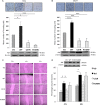
Invasive and migratory properties of C51R and R140W. 2D Boyden assay chamber cell invasion experiments in TPC1 (A) and MCF7 (B) cells transfected with VO, WT PBF, C51R and R140W mutants, with representative photomicrographs shown above. Data presented as mean values ± s.e. (n = 2). (C) Representative classical scratch wound assays in NIH 3T3 cells stably transfected with VO, WT PBF, C51R and R140W substitutions at 4 and 6 h, with percentages of wound recovery shown graphically in D. Inset – Western blotting analysis of PBF levels in the stable NIH 3T3 cells using an anti-PBF antibody. Data presented as mean values ± s.e. (n = 3). *P < 0.05, ***P < 0.001.
Figure 5
Figure 5
PBF mutations C51R and R140W retain the capability to repress radioiodide uptake in vitro. Radioiodide uptake experiments in TPC1 (A) and MCF7 (B) cells transiently transfected with VO, NIS-MYC and VO, WT PBF, C51R or R140W. Uptake was normalised to protein concentration and specific NIS-mediated uptake was demonstrated using the NIS inhibitor sodium perchlorate. Data presented as mean values ± s.e. (n = 3). **P < 0.01, ***P < 0.001. Immunofluorescent microscopy of NIS-MYC (red) with FLAG-tagged WT PBF, C51R or R140W (green), using the NIS and FLAG antibodies, respectively, in HeLa (C) and MCF7 (D) cells. NIS-MYC was observed predominantly at the plasma membrane with VO co-transfection and with the co-transfection of WT PBF, C51R or R140W mislocalised to intracellular vesicles, ER and Golgi, respectively, where co-localisation was observed (yellow). Magnification = 100×. Bars = 20 µm.
Figure 6
Figure 6
C51R and R140W substitutions lose cell transformation ability compared to WT PBF. (A) Colony formation assays in TPC1 cells stably transfected with VO, WT PBF, C51R and R140W; representative photomicrographs shown above. (B) Soft agar assays in NIH 3T3 cells stably transfected with VO, WT PBF, C51R and R140W mutants. Representative images above illustrate relative numbers of colonies. Inset – Western blots of PBF expression in the stable cell lines using an anti-PBF antibody. Data presented as mean values ± s.e. (n = 3, each with >3 replicates). *P < 0.05.

References

    1. Boelaert K, McCabe CJ, Tannahill LA, Gittoes NJ, Holder RL, Watkinson JC, Bradwell AR, Sheppard MC, Franklyn JA. 2003. Pituitary tumor transforming gene and fibroblast growth factor-2 expression: potential prognostic indicators in differentiated thyroid cancer. Journal of Clinical Endocrinology and Metabolism 88 2341–2347. (10.1210/jc.2002-021113) - DOI - PubMed
    1. Boelaert K, Smith VE, Stratford AL, Kogai T, Tannahill LA, Watkinson JC, Eggo MC, Franklyn JA, McCabe CJ. 2007. PTTG and PBF repress the human sodium iodide symporter. Oncogene 26 4344–4356. (10.1038/sj.onc.1210221) - DOI - PubMed
    1. Cerami E, Gao J, Dogrusoz U, Gross BE, Sumer SO, Aksoy BA, Jacobsen A, Byrne CJ, Heuer ML, Larsson E, et al. 2012. The cBio cancer genomics portal: an open platform for exploring multidimensional cancer genomics data. Cancer Discovery 2 401–404. (10.1158/2159-8290.CD-12-0095) - DOI - PMC - PubMed
    1. Chin L, Andersen JN, Futreal PA. 2011. Cancer genomics: from discovery science to personalized medicine. Nature Medicine 17 297–303. (10.1038/nm.2323) - DOI - PubMed
    1. Forbes SA, Bhamra G, Bamford S, Dawson E, Kok C, Clements J, Menzies A, Teague JW, Futreal PA, Stratton MR. 2008. The Catalogue of Somatic Mutations in Cancer (COSMIC). Current Protocols in Human Genetics Chapter 10 Unit 10 11. (10.1002/0471142905.hg1011s57) - DOI - PMC - PubMed

LinkOut - more resources