

Influence of longitudinal radiation exposure from microcomputed tomography scanning on skeletal muscle function and metabolic activity in female CD-1 mice
- PMID: 28676556
- PMCID: PMC5506525
- DOI: 10.14814/phy2.13338
Influence of longitudinal radiation exposure from microcomputed tomography scanning on skeletal muscle function and metabolic activity in female CD-1 mice
Abstract
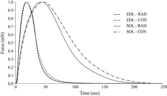
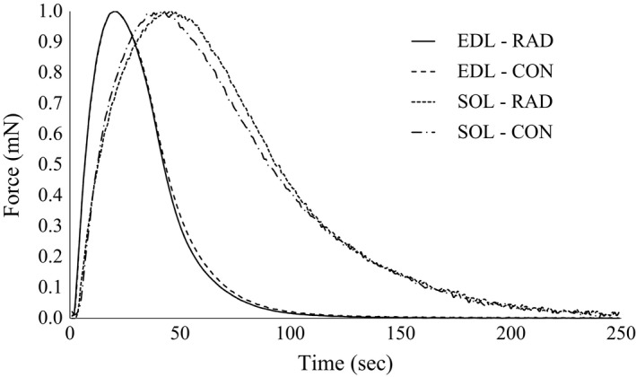
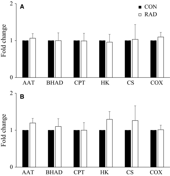
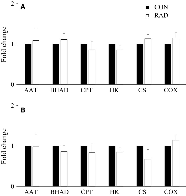
Microcomputed tomography (μCT) is an imaging technology to assess bone microarchitecture, a determinant of bone strength. When measured in vivo, μCT exposes the skeletal site of interest to a dose of radiation, in addition to nearby skeletal muscles as well. Therefore, the aim of this study was to determine the effects of repeated radiation exposure from in vivo μCT on muscle health - specifically, muscle morphometrics, contractile function, and enzyme activity. This study exposed the right hind limb of female mice to either a low (26 cGy) or moderate (46 cGy) dose, at 2, 4, and 6 months of age, while the left hind limb of the same animal was exposed to a single dose at 6 months to serve as a nonirradiated control. Muscle weight, cross-sectional area, isometric contractile function, and representative maximal enzyme activities of amino acid, fatty acid, glucose, and oxidative metabolism in extensor digitorum longus (EDL) and soleus were assessed. Low-dose radiation had no effect. In contrast, moderate-dose radiation resulted in a 5% increase in time-to-peak tension and 16% increase in half-relaxation time of isometric twitches in EDL, although these changes were not seen when normalized to force. Moderate-dose radiation also resulted in an ~33% decrease in citrate synthase activity in soleus but not EDL, with no changes to the other enzymes measured. Thus, three low doses of radiation over 6 months had no effect on contractile function or metabolic enzyme activity in soleus and EDL of female mice. In contrast, three moderate doses of radiation over 6 months induced some effects on metabolic enzyme activity in soleus but not EDL Future studies that wish to investigate muscle tissue that is adjacent to scanned bone should take radiation exposure dose into consideration.
Keywords: Citrate synthase; X‐ray; extensor digitorum longus; soleus.
© 2017 The Authors. Physiological Reports published by Wiley Periodicals, Inc. on behalf of The Physiological Society and the American Physiological Society.
Figures
Similar articles
-
Contraction parameters, myosin composition and metabolic enzymes of the skeletal muscles of the etruscan shrew Suncus etruscus and of the common European white-toothed shrew Crocidura russula (Insectivora: soricidae).J Exp Biol. 1999 Sep;202(Pt 18):2461-73. doi: 10.1242/jeb.202.18.2461. J Exp Biol. 1999. PMID: 10460733
-
Endurance training adaptations modulate the redox-force relationship of rat isolated slow-twitch skeletal muscles.Clin Exp Pharmacol Physiol. 2003 Jan-Feb;30(1-2):77-81. doi: 10.1046/j.1440-1681.2002.03794.x. Clin Exp Pharmacol Physiol. 2003. PMID: 12542458
-
The effects of dietary creatine supplements on the contractile properties of rat soleus and extensor digitorum longus muscles.Exp Physiol. 2001 Mar;86(2):185-90. doi: 10.1113/eph8602131. Exp Physiol. 2001. PMID: 11429633
-
Influence of ageing on the fatigability of isolated mouse skeletal muscles from mature and aged mice.Exp Physiol. 2002 Jan;87(1):77-82. doi: 10.1113/eph8702224. Exp Physiol. 2002. PMID: 11805861
-
The effect of obesity on the contractile performance of isolated mouse soleus, EDL, and diaphragm muscles.J Appl Physiol (1985). 2017 Jan 1;122(1):170-181. doi: 10.1152/japplphysiol.00836.2016. Epub 2016 Nov 17. J Appl Physiol (1985). 2017. PMID: 27856719
Cited by
-
The Influence of Supplemental Dietary Linoleic Acid on Skeletal Muscle Contractile Function in a Rodent Model of Barth Syndrome.Front Physiol. 2021 Aug 19;12:731961. doi: 10.3389/fphys.2021.731961. eCollection 2021. Front Physiol. 2021. PMID: 34489741 Free PMC article.
References
-
- Alhomida, A. S. 2001. Evaluation of theophylline‐stimulated changes in carnitine palmitoyltransferase activity in skeletal muscle and liver of rats. J. Enzym. Inhib. 16:177–183. - PubMed
-
- Bandstra, E. R. , Thompson R. W., Nelson G. A., Willey J. S., Judex S., Cairns M. A., et al. 2009. Musculoskeletal changes in mice from 20–50 cGy of simulated galactic cosmic rays. Radiat. Res. 172:21–29. - PubMed
-
- Barclay, C. J. 2005. Modelling diffusive O(2) supply to isolated preparations of mammalian skeletal and cardiac muscle. J. Muscle Res. Cell Motil. 26:225–235. - PubMed
-
- Bass, A. , Brdiczka D., Eyer P., Hofer S., and Pette D.. 1969. Metabolic differentiation of distinct muscle types at the level of enzymatic organization. Eur. J. Biochem. 10:198–206. - PubMed
MeSH terms
Substances
LinkOut - more resources
Full Text Sources
Other Literature Sources
