. 2017 Jul 4;7(1):4571.
doi: 10.1038/s41598-017-04870-8.

Human neutrophils phagocytose and kill Acinetobacter baumannii and A. pittii

Affiliations

Human neutrophils phagocytose and kill Acinetobacter baumannii and A. pittii

María Lázaro-Díez et al. Sci Rep. .

Erratum in

Abstract

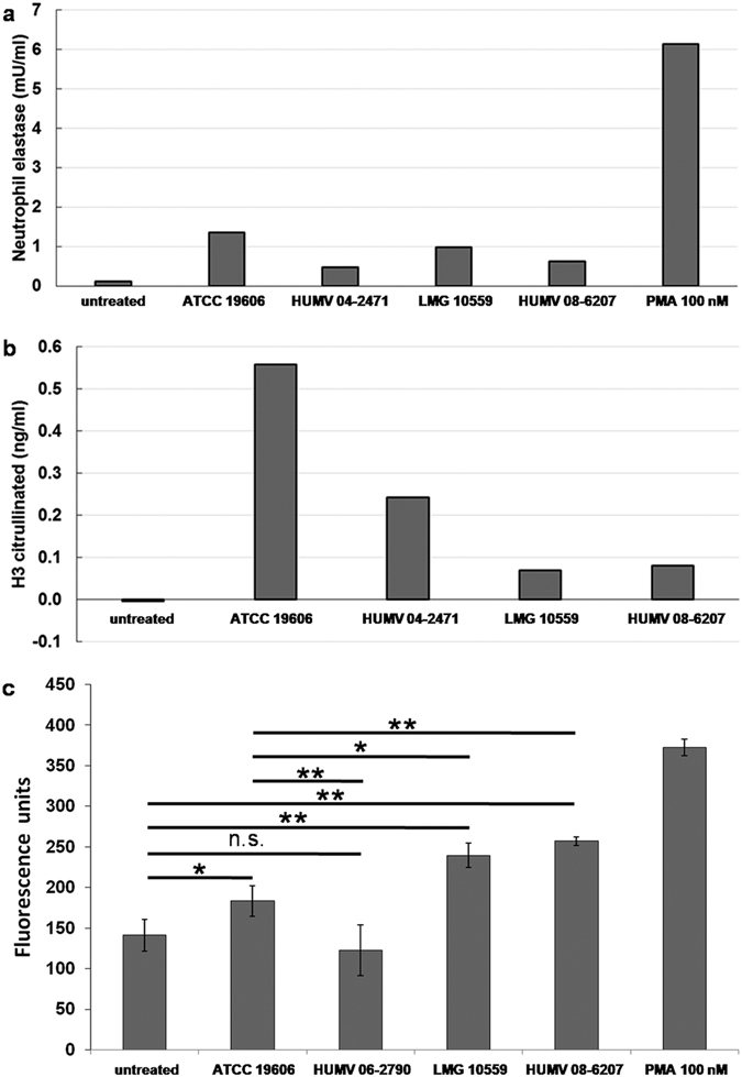
Acinetobacter baumannii is a common cause of health care associated infections worldwide. A. pittii is an opportunistic pathogen also frequently isolated from Acinetobacter infections other than those from A. baumannii. Knowledge of Acinetobacter virulence factors and their role in pathogenesis is scarce. Also, there are no detailed published reports on the interactions between A. pittii and human phagocytic cells. Using confocal laser and scanning electron microscopy, immunofluorescence, and live-cell imaging, our study shows that immediately after bacteria-cell contact, neutrophils rapidly and continuously engulf and kill bacteria during at least 4 hours of infection in vitro. After 3 h of infection, neutrophils start to release neutrophil extracellular traps (NETs) against Acinetobacter. DNA in NETs colocalizes well with human histone H3 and with the specific neutrophil elastase. We have observed that human neutrophils use large filopodia as cellular tentacles to sense local environment but also to detect and retain bacteria during phagocytosis. Furthermore, co-cultivation of neutrophils with human differentiated macrophages before infections shows that human neutrophils, but not macrophages, are key immune cells to control Acinetobacter. Although macrophages were largely activated by both bacterial species, they lack the phagocytic activity demonstrated by neutrophils.


Conflict of interest statement

The authors declare that they have no competing interests.

Figures

Figure 1
Figure 1
Contact and phagocytosis of Acinetobacter by human neutrophils. Human neutrophils were infected for 30 min (a), 60 min (b,c) or 2 h (d–f) with A. baumannii ATCC 19606T, fixed and processed for immunofluorescence labelling. Bacteria were detected with anti-A. baumannii rabbit antibody (red). Actin cytoskeleton was labelled with Atto 488 phalloidin (green) and nuclei are stained with DAPI (blue) (a–c,f). (a) Single stack; (b and d–f) maximal projections; (c) cross-sectional view. Arrow in (b) indicates a pseudopod in close contact with a bacterium. In (d,e) double-immunofluorescence images show extracellular bacteria (green), debris of intracellular bacteria (red) and bacterial and cellular DNA (blue). (f) As control, fresh untreated neutrophils were incubated in parallel during 4 h. Micrographs were originally captured at ×400 magnification (a,f) or ×600 magnification (b–e). Scale bars, (a,f) 5 µm; (b,d,e) 2 µm.
Figure 2
Figure 2
Live/Dead staining in unfixed neutrophils. Neutrophils were infected with Acinetobacter strains, then exposed to components of the live/dead kit, propidium iodide and SYTO9. Upper panels: in merged images, live bacteria appear green, dead bacteria appear red and eukaryotic nuclei appear pink. Lower panels show selected z-stacks at high magnifications (red channel) of the boxed areas in the upper panel. Untreated similarly stained cells served as control (C). Original magnifications: upper panels ×400; lower panels ×1000. Scale bars, 5 µm.
Figure 3
Figure 3
Capture and phagocytosis of Acinetobacter by human neutrophils. Pictures show SEM microphotographs (a,c,d,e) or immunofluorescence (b) images of infected neutrophils (3 h, strain ATCC 19606T). Large filopodia were observed in infected cultures in close contact with bacteria (b,c). Some of these filopodia completely surround two bacteria (asterisks in c) while pseudopods are catching bacteria attached to the inert surface (arrows in c). In (b) bacteria were detected with anti-A. baumannii rabbit antibody (red), actin cytoskeleton was labelled with Atto 488 phalloidin (green) and nuclei were stained with DAPI (blue). Unstimulated neutrophils show round shapes (d). (e) Detail of the boxed area in (d) Micrographs were originally captured at ×4000 (a), ×600 (b), ×10000 (c), ×500 (d) or ×9000 magnification (e). Scale bars, (a) 10 µm; (b,c,e) 5 µm; (d) 100 µm.
Figure 4
Figure 4
NETs production by human neutrophils infected with Acinetobacter. Pictures show a SEM microphotograph (a) or immunofluorescence images (b–e) of neutrophils 4 h post-infection: (a,c–e) strain ATCC 19606T; (b) strain HUMV 06-2790. From (b to e) bacteria were detected with anti-A. baumannii rabbit antibody (red), actin was labelled with Atto 488 phalloidin (green), and DNA was stained with DAPI (blue). Micrographs were originally captured at ×15000 (a), ×600 (b,d), ×400 (c) or ×200 magnification (e). Scale bars, (a–d) 5 µm; (e) 100 µm.
Figure 5
Figure 5
Quantification of elastase, citrullinated histone H3 and extracellular DNA using SYTOX Green. (a) Measurement of released neutrophil elastase. Human neutrophils were infected with Acinetobacter strains or treated with PMA for four hours, washed, and treated with S7 nuclease for 15 min. The supernatant from each well was assayed. Samples were tested in triplicate. (b) Measurement of citrullinated histone H3 (CitH3). Human neutrophils were infected with Acinetobacter strains for 4 hours. Supernatants were centrifuged to remove cellular debris and then tested in the ELISA. Samples were tested in triplicate. The concentrations of total neutrophil elastase and CitH3 in the analyzed samples were estimated from standard curves obtained for each assay. (c) Quantification of fluorescence after infection experiments using SYTOX Green. Supernatants from unstained infected cultures were partially digested with DNAse I and stained with SYTOX Green. Each bar indicates the average of three independent experiments ± SD. Asterisks indicate: *p = 0.0004; **p < 0.00001; n.s., p = 0.1610.

References

    1. Clark NM, Zhanel GG, Lynch JP., 3rd Emergence of antimicrobial resistance among Acinetobacter species: a global threat. Curr Opin Crit Care. 2016;22:491–499. doi: 10.1097/MCC.0000000000000337. - DOI - PubMed
    1. Lahmer T, et al. Acinetobacter baumannii sepsis is fatal in medical intensive care unit patients: six cases and review of literature. Anaesth Intensive Care. 2014;42:666–668. - PubMed
    1. Greene C, Vadlamudi G, Newton D, Foxman B, Xi C. The influence of biofilm formation and multidrug resistance on environmental survival of clinical and environmental isolates of Acinetobacter baumannii. Am J Infect Control. 2016;44:e65–71. doi: 10.1016/j.ajic.2015.12.012. - DOI - PMC - PubMed
    1. Espinal P, Marti S, Vila J. Effect of biofilm formation on the survival of Acinetobacter baumannii on dry surfaces. J Hosp Infect. 2012;80:56–60. doi: 10.1016/j.jhin.2011.08.013. - DOI - PubMed
    1. Vila-Farres X, et al. In vitro activity of several antimicrobial peptides against colistin-susceptible and colistin-resistant Acinetobacter baumannii. Clin Microbiol Infect. 2012;18:383–387. doi: 10.1111/j.1469-0691.2011.03581.x. - DOI - PubMed

Publication types

MeSH terms