

Lower In vivo Myo-Inositol in the Anterior Cingulate Cortex Correlates with Delayed Melatonin Rhythms in Young Persons with Depression
- PMID: 28676736
- PMCID: PMC5476733
- DOI: 10.3389/fnins.2017.00336
Lower In vivo Myo-Inositol in the Anterior Cingulate Cortex Correlates with Delayed Melatonin Rhythms in Young Persons with Depression
Abstract
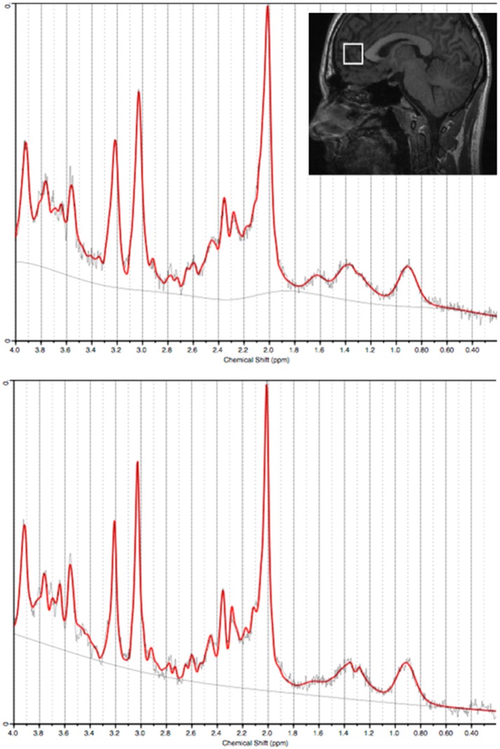
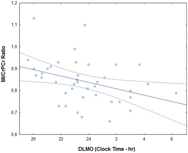
Myo-inositol, a second messenger glucose isomer and glial marker, is potentiated by melatonin. In addition to common abnormalities in melatonin regulation, depressive disorders have been associated with reduced myo-inositol in frontal structures. This study examined associations between myo-inositol in the anterior cingulate cortex and the timing of evening melatonin release. Forty young persons with unipolar depression were recruited from specialized mental health services (20.3 ± 3.8 years old). Healthy controls were recruited from the community (21.7 ± 2.6 years old). The timing of dim light melatonin onset (DLMO) was estimated using salivary melatonin sampling. Myo-inositol concentrations (MI/CrPCr ratio) in the anterior cingulate cortex were obtained using proton magnetic resonance spectroscopy. After controlling for age, sex, and CrPCr concentration the depression group had significantly lower MI/CrPCr ratios than healthy controls [F(4, 75) = 11.4, p = 0.001]. In the depression group, later DLMO correlated with lower MI/CrPCr ratio (r = -0.48, p = 0.014). These findings suggest that neurochemical changes in the frontal cortex are associated with circadian disruptions in young persons with depression.
Keywords: circadian; depression; magnetic resonance spectroscopy; melatonin; myo-inositol.
Figures
References
-
- American Psychiatric Association (2000). Diagnostic and Statistical Manual of Mental Disorders, 4th Revised Edn. Washington, DC: American Psychiatric.
LinkOut - more resources
Full Text Sources
Other Literature Sources