Review
. 2017 Jun 20:7:276.
doi: 10.3389/fcimb.2017.00276. eCollection 2017.

Neuroinflammation and Infection: Molecular Mechanisms Associated with Dysfunction of Neurovascular Unit

Affiliations
Review

Neuroinflammation and Infection: Molecular Mechanisms Associated with Dysfunction of Neurovascular Unit

Abolghasem Tohidpour et al. Front Cell Infect Microbiol. .

Abstract

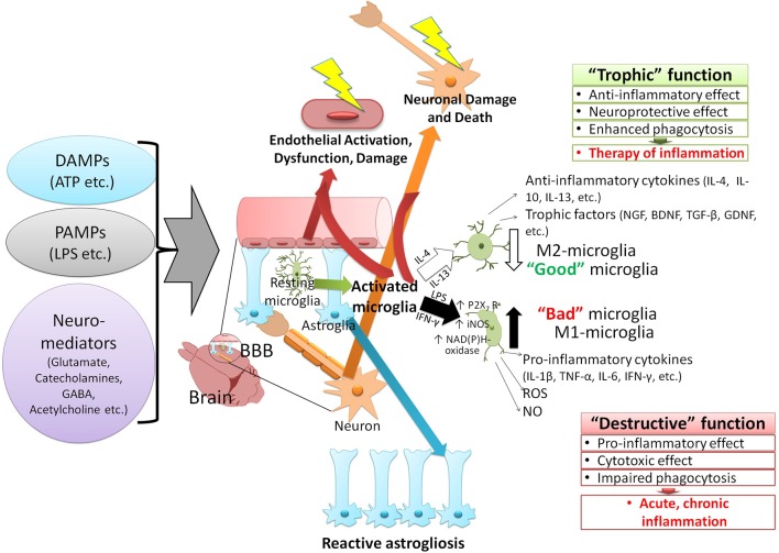
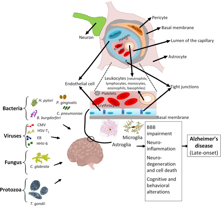
Neuroinflammation is a complex inflammatory process in the central nervous system, which is sought to play an important defensive role against various pathogens, toxins or factors that induce neurodegeneration. The onset of neurodegenerative diseases and various microbial infections are counted as stimuli that can challenge the host immune system and trigger the development of neuroinflammation. The homeostatic nature of neuroinflammation is essential to maintain the neuroplasticity. Neuroinflammation is regulated by the activity of neuronal, glial, and endothelial cells within the neurovascular unit, which serves as a "platform" for the coordinated action of pro- and anti-inflammatory mechanisms. Production of inflammatory mediators (cytokines, chemokines, reactive oxygen species) by brain resident cells or cells migrating from the peripheral blood, results in the impairment of blood-brain barrier integrity, thereby further affecting the course of local inflammation. In this review, we analyzed the most recent data on the central nervous system inflammation and focused on major mechanisms of neurovascular unit dysfunction caused by neuroinflammation and infections.

Keywords: blood-brain barrier; brain development; immune response; infectious diseases; neurodegeneration.


Figures

Figure 1
Figure 1
The paradigm of the CNS neuroinflammation. Various factors can activate the immune response of the CNS and induce neuroinflammation. These stimuli are classified into two groups: 1—pathogen-associated molecular patterns (PAMPs), which are produced by the invading microorganisms of the CNS and 2—damage-associated molecular patterns (DAMPs), molecules that are released by host due to onset of traumatic conditions or interaction with some neurotransmitters (i.e., glutamate, GABA, and acetylcholine). The immune responses to the CNS stimuli vary based on the type of stimulation but generally lead to similar outcomes such as immune adaptation, dysfunction, degeneration, and resolution. Activation of the resting microglia and converting them to two distinct phenotypes depends on various cytokines produced by surrounding cells (glia, neurons, migratory immune cells). The release of interleukins 4 and 13 (IL-4, IL-13) gives rise to M1 phenotype (anti-inflammatory) of microglia, which express inflammatory cytokines (interleukin 4, 10, and 13), cell growth factors (i.e., NGF, BDNF, TGF-β, GDNF), and exert anti-inflammatory effects. Interferon-γ (IFN-γ) and the lipopolysaccharide (LPS) of bacteria, on the other hand, activate the M2 phenotype (pro-inflammatory) of microglia. The M2 phenotype is characterized by 1—activation of purinergic receptors P2X7 subtype (activated by ATP, promoting the inflammation and destruction of cells by forming channels and pores), and 2—expression of enzymes which generate reactive oxygen and nitrogen [NAD(P)H-oxidase, iNOS], and trigger the expression of proinflammatory cytokines (IL-1β, TNF-α, IL-6, IFN- γ). Activation of microglia, especially the formation of M2 phenotype exacerbates the damage to BBB (in particular neurons and endothelial cells). Effects of these agents (PAMPs, DAMPs, neuromediators) on astroglia cause their proliferation, activation (reactive astrogliosis), and dysfunction (in particular, increased procoagulant activity and thrombosis). These CNS stimuli also cause endothelial injury, damage, and neuronal death. GABA, γ-aminobutyric acid; IL, Interleukin; NGF, nerve growth factor; BDNF, brain-derived neurotrophic factor; TGF, transforming growth factor; GDNF, glial-derived neurotrophic factors; iNOS, inducible nitric oxide synthase; TNF, tumor necrosis factor; IFN, interferon; ROS, reactive oxygen species; NO, nitric oxide.
Figure 2
Figure 2
Association of infectious agents with Alzheimer's disease. Chronic infections caused by major infectious agents, i.e., Helicobacter pylori, various types of spirochetes, including periodontal pathogen spirochetes and Borrelia burgdorferi, Porphyromonas gingivalis, Chlamydophila pneumoniae, Cytomegalovirus, Herpes simplex virus type 1, Epstein-Bar virus, Human herpes virus 6, Candida glabrata and Toxoplasma gondii are associated with development of AD. Early life exposure to these pathogenic agents can activate the resting microglia and astroglia, trigger the migration of immune cells to the neuro-endothelial tissue, degrade cell-cell tight junctions, and cause the breakdown of BBB. These activities result in development of various side effects such as neuronal damage, neuroinflammation and ultimately predispose the adult patient to develop AD.

Similar articles

Cited by

References

    1. Agapitov A. V., Haynes W. G. (2002). Role of endothelin in cardiovascular disease. J. Renin Angiotensin Aldosterone Syst. 3, 1–15. 10.3317/jraas.2002.001 - DOI - PubMed
    1. Agostini S., Clerici M., Mancuso R. (2014). How plausible is a link between HSV-1 infection and Alzheimer's disease? Expert Rev. Anti Infect. Ther. 12, 275–278. 10.1586/14787210.2014.887442 - DOI - PubMed
    1. Ajami B., Bennett J. L., Krieger C., Tetzlaff W., Rossi F. M. (2007). Local self-renewal can sustain CNS microglia maintenance and function throughout adult life. Nat. Neurosci. 10, 1538–1543. 10.1038/nn2014 - DOI - PubMed
    1. Alexander C., Rietschel E. T. (2001). Bacterial lipopolysaccharides and innate immunity. J. Endotoxin Res. 7, 167–202. 10.1179/096805101101532675 - DOI - PubMed
    1. Alfonso-Loeches S., Ureña-Peralta J., Morillo-Bargues M. J., Gómez-Pinedo U., Guerri C. (2016). Ethanol-induced TLR4/NLRP3 neuroinflammatory response in microglial cells promotes leukocyte infiltration across the BBB. Neurochem. Res. 41, 193–209. 10.1007/s11064-015-1760-5 - DOI - PubMed

Publication types

MeSH terms