. 2017:Suppl 218:5-13.
doi: 10.2340/00015555-2717. Epub 2017 Jul 5.

Innovations and Developments in Dermatologic Non-invasive Optical Imaging and Potential Clinical Applications

Affiliations

Innovations and Developments in Dermatologic Non-invasive Optical Imaging and Potential Clinical Applications

Eric Tkaczyk. Acta Derm Venereol. 2017.

Abstract

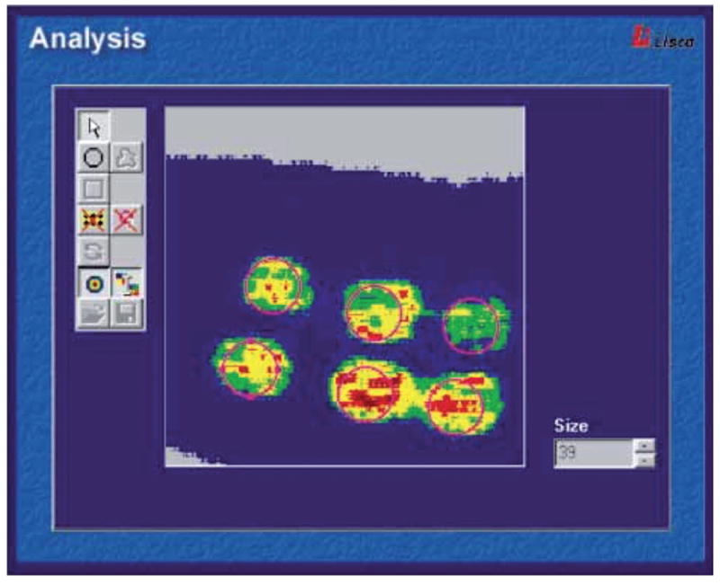
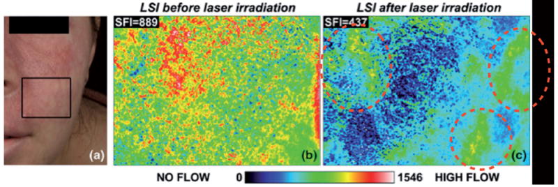
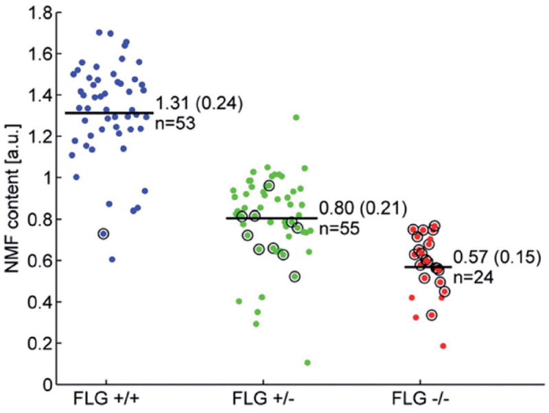
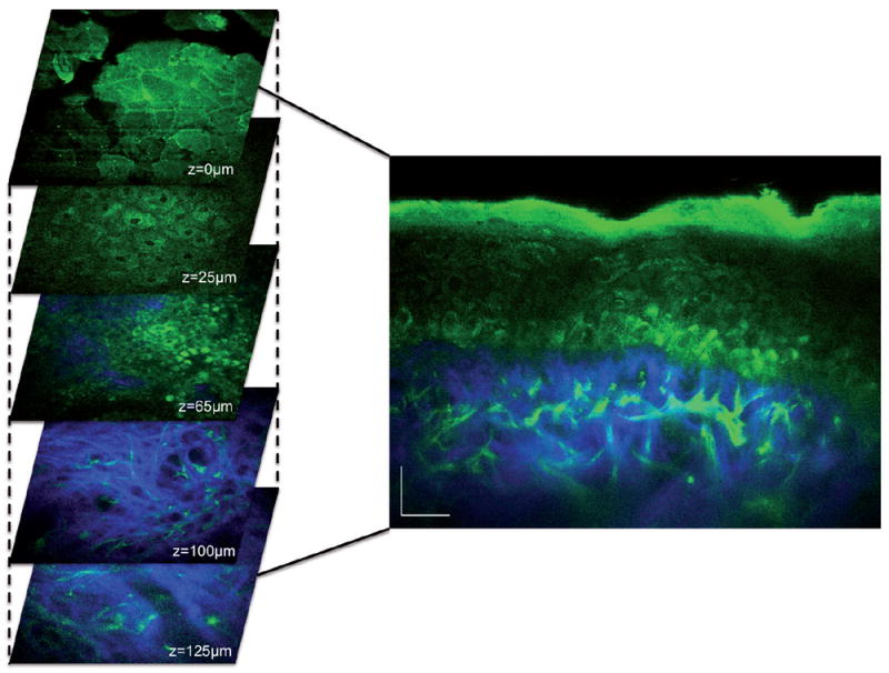
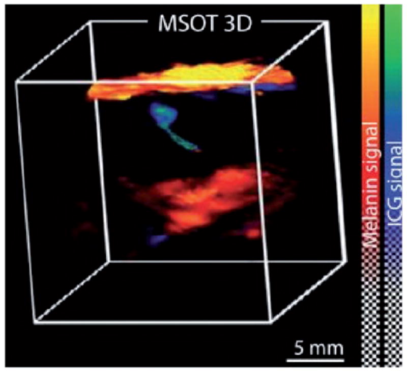
Most dermatologists are aware of the benefits of dermoscopy, and a few are familiar with laser-scanning confocal microscopy. Beyond confocal, there are fully 11 different categories of optical techniques that have been applied to clinical dermatology. This article first provides a comprehensive tabular overview of all these optical diagnostic technologies and then details 4 of the lesser known innovations that are already available or still in development (laser Doppler and speckle imaging, Raman spectroscopy, multiphoton microscopy, photoacoustic tomography), with some potential applications in clinical dermatology (blood flow monitoring, skin cancer diagnosis, composition measurements in atopic dermatitis, skin rejuvenation measurement, and noninvasive sentinel lymph node assessment in melanoma). These methods present many advantages, being non-invasive, portable, and rapid. The development of optics in biological and biomedical sciences (i.e. biophotonics) requires not only deep insight into the applications but also synergistic collaboration be-tween engineers and clinicians.


Conflict of interest statement

The author declares no conflicts of interest.

Figures

Fig. 1
Fig. 1. Laser doppler imaging of blood flow in a patch test
Printed with permission from Fullerton et al. (2002) (10). The perfusion image can be analysed by an integrated system software. The relative colour scale extends from the smallest and the largest perfusion value (from green to red). (https://www.perimed-instruments.com/skin-patch-testing).
Fig. 2
Fig. 2. Laser Speckle Imaging (LSI) of a Caucasian female patient with a port wine stain involving the V2 dermatomal distribution
Printed with permission from Huang Y.C. et al. (2008) (11). (a) Photograph. (b) Speckle Flow Index images taken from the marked region of interest immediately before and (c) 15 min after laser therapy. Colour range indicates the level of blood flow in this area.
Fig. 3
Fig. 3
Cloud plot of natural moisturising factor (NMF) values in newborns, obtained from Raman spectra and categorized by filaggrin (FLG) genotype (final genotype after full screening: FLG+/+, FLG+/–, FLG−/−). From O’Regan GM et al. (2010) (15). For each group, the number of patients and NMF level (mean ± SD) are indicated in the figure. a.u.: arbitrary units.
Fig. 4
Fig. 4. In vivo MPM imaging of normal human skin
Left, horizontal sections of MPM images (x–y scans) at different depths showing images of: the stratum corneum (z = 0 μm), keratinocytes normally distributed in the stratum spinosum (z=25 μm), basal cells (green) surrounding dermal papilla (blue; z=65 μm), collagen (blue) and elastin fibers (green) in the dermis (z=100 μm;). Right, cross-sectional view (x–z scan) corresponding to a vertical plane through the horizontal sections on the left. Scale bar is 20 μm. Image kindly provided by Dr Mihaela Balu from University of California, Irvine/Beckman Laser Institute.
Fig. 5
Fig. 5. Preoperative assessment of sentinel lymph node melanin content using multispectral opto-acoustic tomography (MSOT)
Printed with permission from Stoffels I. et al. (2015), (30). Sentinel lymph node of a patient with metastasis per a combined 3D rendering of an MSOT image taken by the 3D detector that shows both melanin (red) and indocyanine green (ICG) localization. The skin pigment appears in yellow. MSOT imaging was able to localize sentinel lymph node and melanin to provide information on the metastatic status of the lymph node.

References

    1. Tkaczyk T, Xu C. Introduction to the bio-optics: design and application. Biomed Opt Express. 2015;6:4899–4900. - PMC - PubMed
    1. Sharif SA, Taydas E, Mazhar A, Rahimian R, Kelly KM, Choi B, et al. Noninvasive clinical assessment of port-wine stain birthmarks using current and future optical imaging technology: a review. Br J Dermatol. 2012;167:1215–1223. - PMC - PubMed
    1. Garbe C, Peris K, Hauschild A, Saiag P, Middleton M, Bastholt L, et al. Diagnosis and treatment of melanoma. European consensus-based interdisciplinary guideline – Update 2016. Eur J Cancer. 2016;63:201–217. - PubMed
    1. Stratigos A, Garbe C, Lebbe C, Malvehy J, del Marmol V, Pehamberger H, et al. Diagnosis and treatment of invasive squamous cell carcinoma of the skin: European consensus-based interdisciplinary guideline. Eur J Cancer. 2015;51:1989–2007. - PubMed
    1. Trakatelli M, Morton C, Nagore E, Ulrich C, Del Marmol V, Peris K, et al. Update of the European guidelines for basal cell carcinoma management. Eur J Dermatol. 2014;24:312–329. - PubMed

LinkOut - more resources