. 2017 Jul 4;22(7):1106.
doi: 10.3390/molecules22071106.

Newly Synthesized Doxorubicin Complexes with Selected Metals-Synthesis, Structure and Anti-Breast Cancer Activity

Affiliations

Newly Synthesized Doxorubicin Complexes with Selected Metals-Synthesis, Structure and Anti-Breast Cancer Activity

Agata Jabłońska-Trypuć et al. Molecules. .

Abstract

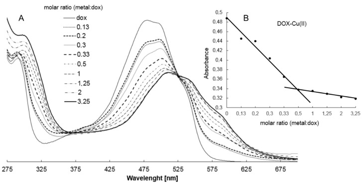
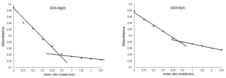
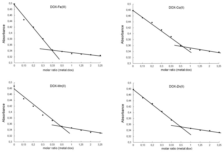
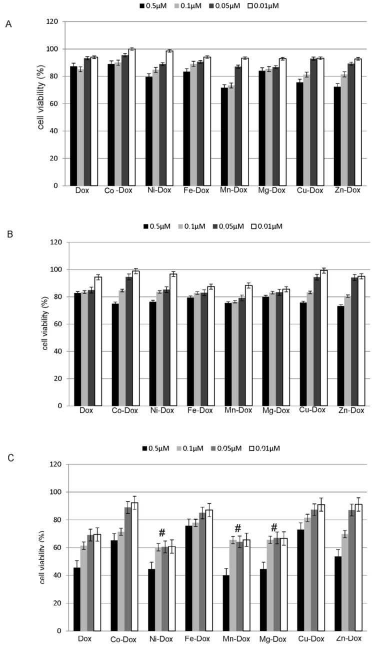
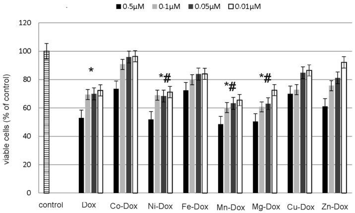
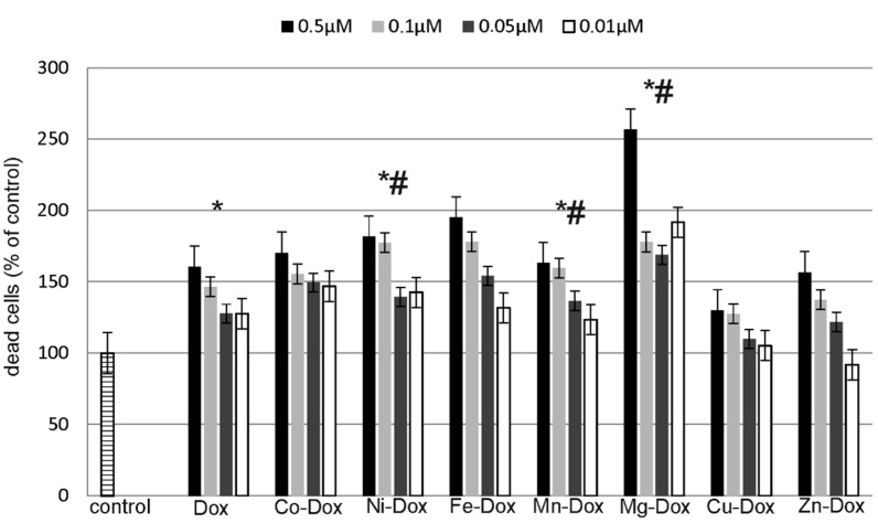
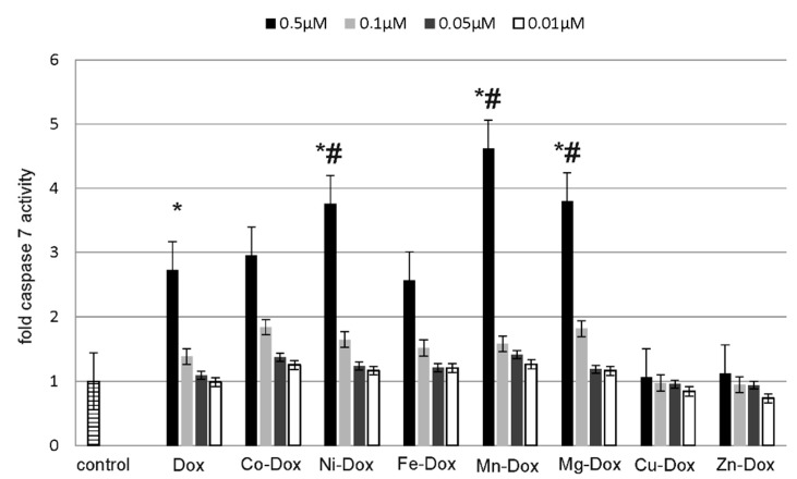
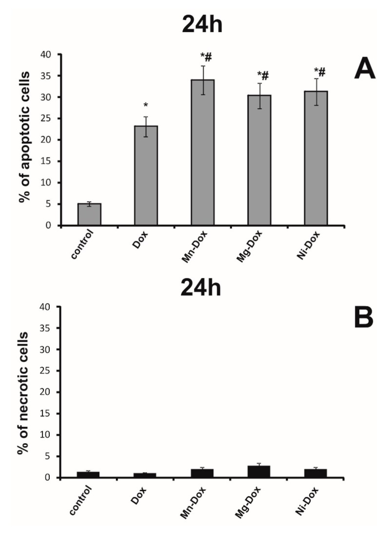
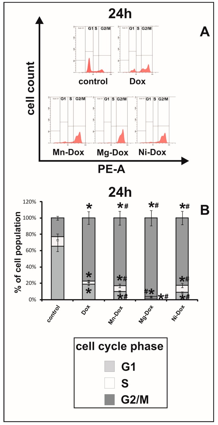
Doxorubicin (DOX) is very effective chemotherapeutic agent, however it has several major drawbacks. Therefore the motivation for developing novel drug complexes as anticancer agents with different mechanism of action has arisen. The aim of the present study was to evaluate the influence of newly synthesized DOX complexes with selected metals (Mg, Mn, Co, Ni, Fe, Cu, Zn) on apoptosis, cell cycle, viability, proliferation and cytotoxicity in the breast cancer cell line MCF-7. Complexation of DOX with metals has likewise been the subject of our research. The current work showed that the tested bivalent metals at a given pH condition formed metal:DOX complexes in a ratio of 2:1, while iron complexes with DOX in a ratio of 3:1. The studies also showed that selected metal-DOX complexes (Mg-DOX, Mn-DOX, Ni-DOX) at 0.5 µM concentration significantly decreased cell viability and proliferation, however they increased caspase 7 activity. Results also indicated that studied metal-DOX complexes showed high cytotoxicity in MCF-7 cells. Therefore they were chosen for cell cycle check-points and apoptosis/necrosis analysis studied by flow cytometry. Obtained results suggest that doxorubicin complexed by specified metals can be considered as a potential anti-breast cancer agent, which is characterized by a higher efficacy than a parent drug.

Keywords: MCF-7; breast cancer; doxorubicin; metals.


Conflict of interest statement

The authors declare no conflict of interest.

Figures

Figure 1
Figure 1
Structural formula of the selected anthracyclines regarding the functional groups of the individual compounds.
Figure 2
Figure 2
IR KBr (a) and ATR (b) and Raman (c) spectra of DOX (continuous lines) and IR KBr spectra of Cu-DOX complex (the dotted line) (d).
Figure 3
Figure 3
(A) Absorption spectra of DOX in the presence of various amounts of Cu(II) in Tris-HCl buffer pH 7.01 (different types of lines are labeled with a molar ratio Cu(II):DOX 0.13–2.0); (B) absorbance as a function of [Cu(II)]:[DOX] molar ratio at 500 nm.
Figure 4
Figure 4
Absorbance as a function of [Metal]:[DOX] molar ratio at 500 nm. Metal-Dox complexes are water-soluble and they were prepared in Tris-HCl buffer pH 7.01.
Figure 4
Figure 4
Absorbance as a function of [Metal]:[DOX] molar ratio at 500 nm. Metal-Dox complexes are water-soluble and they were prepared in Tris-HCl buffer pH 7.01.
Figure 5
Figure 5
Proposal structure of complex metal-DOX.
Figure 6
Figure 6
The effect of DOX, Cu-DOX, Zn-DOX, Co-DOX, Ni-DOX, Fe-DOX, Mn-DOX and Mg-DOX on cell proliferation of MCF-7 cells. The cells were incubated with 0.5 µM, 0.1 µM, 0.05 µM and 0.01 µM DOX and Metal-DOX complexes for 2 h (a), 4 h (b) and 24 h (c). Mean values from five independent experiments ± SD are shown. Significant alterations are expressed relative to DOX—controls and marked with crosses (#). Statistical significance was considered if # p < 0.05.
Figure 7
Figure 7
Co, Ni, Fe, Mn, Mg, Cu and Zn cytotoxicity in MCF-7 cells. The cells were incubated with 0.25 µM, 0.05 µM, 0.025 µM and 0.0005 µM of metal chlorides for 24 h. Mean values from five independent experiments ± SD are shown. Results are statistically insignificant.
Figure 8
Figure 8
The effect of DOX, Cu-DOX, Zn-DOX, Co-DOX, Ni-DOX, Fe-DOX, Mn-DOX and Mg-DOX on cell viability of MCF-7 cells. The cells were incubated with 0.5 µM, 0.1 µM, 0.05 µM and 0.01 µM DOX and Metal-DOX complexes for 24 h. Mean values from five independent experiments ± SD are shown. Significant alterations are expressed relative to control untreated cells (marked with asterisks *) and to DOX—controls (marked with crosses #). Statistical significance was considered if *# p < 0.05.
Figure 9
Figure 9
DOX, Cu-DOX, Zn-DOX, Co-DOX, Ni-DOX, Fe-DOX, Mn-DOX and Mg-DOX cytotoxicity in MCF-7 cells. The cells were incubated with 0.5 µM, 0.1 µM, 0.05 µM and 0.01 µM DOX and Metal-DOX complexes for 24 h. Mean values from five independent experiments ± SD are shown. Significant alterations are expressed relative to control untreated cells (marked with asterisks *) and to DOX—controls (marked with crosses #). Statistical significance was considered if #* p < 0.05.
Figure 10
Figure 10
The effect of DOX, Cu-DOX, Zn-DOX, Co-DOX, Ni-DOX, Fe-DOX, Mn-DOX and Mg-DOX on apoptosis in MCF-7 cells. The cells were incubated with 0.5 µM, 0.1 µM, 0.05 µM and 0.01 µM DOX and Metal-DOX complexes for 24 h. Mean values from five independent experiments ± SD are shown. Significant alterations are expressed relative to control untreated cells (marked with asterisks *) and to DOX—controls (marked with crosses #). Statistical significance was considered if *# p < 0.05.
Figure 11
Figure 11
The effect of DOX, Ni-DOX, Mn-DOX and Mg-Dox on apoptosis of MCF-7 cells. The cells were incubated with 0.5 μM DOX, Ni-DOX, Mn-DOX and Mg-DOX for 24 h. Bar graphs presenting the percentage of apoptotic MCF-7 (A) and necrotic MCF-7 (B) cells, are demonstrated. Mean values from five independent experiments ± SD are shown. Significant alterations are expressed relative to control and marked with asterisks (*); significant alterations are expressed relative to DOX-control and marked with crosses (#). Statistical significance was considered if *# p < 0.05.
Figure 12
Figure 12
The effect of DOX, Ni-DOX, Mn-DOX and Mg-DOX on cell cycle distribution of MCF-7 cell line. The cell cycle was measured by propidium iodide staining followed by flow cytometry analysis. Results are shown for cells treated with 0.5 μM Ni-DOX, Mn-DOX and Mg-DOX for 24 h versus untreated controls and versus 0.5 µM DOX. Graphical representation of the cell cycle profiles obtained from flow cytometry measurements in MCF-7 cells is depicted in (A). Bar graph presenting the percentage of cell cycle distribution in MCF-7 after 24 h (B) of treatment with DOX, Ni-DOX, Mn-DOX and Mg-DOX. Significant alterations are expressed relative to control and marked with asterisks (*); significant alterations are expressed relative to DOX-control and marked with crosses (#). Statistical significance was considered if *# p < 0.05.

References

    1. Pilco-Ferreto N., Calaf G.M. Influence of doxorubicin on apoptosis and oxidative stress in breast cancer cell lines. Int. J. Oncol. 2016;49:753–762. doi: 10.3892/ijo.2016.3558. - DOI - PubMed
    1. Turner N., Biganzoli L., Di L.A. Continued value of adjuvant anthracyclines as treatment for early breast cancer. Lancet Oncol. 2015;16:e362–e369. doi: 10.1016/S1470-2045(15)00079-0. - DOI - PubMed
    1. Szuławska A., Czyż M. Molekularne mechanizmy działania antracyklin. Post. Hig. Med. Dośw. 2006;60:78–100. - PubMed
    1. Missailidis S. Anticancer Therapeutics. John Wiley & Sons; Chichester, UK: 2008. pp. 53–54.
    1. Mitry M.A., Edwards J.G. Doxorubicin induced heart failure: Phenotype and molecular mechanisms. IJC Heart Vasc. 2016;10:17–24. doi: 10.1016/j.ijcha.2015.11.004. - DOI - PMC - PubMed

MeSH terms

LinkOut - more resources