. 2017 Jul 5;22(7):1116.
doi: 10.3390/molecules22071116.

Characterization of Danaparoid Complex Extractive Drug by an Orthogonal Analytical Approach

Affiliations

Characterization of Danaparoid Complex Extractive Drug by an Orthogonal Analytical Approach

Cristina Gardini et al. Molecules. .

Abstract

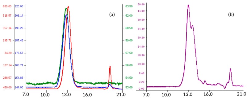
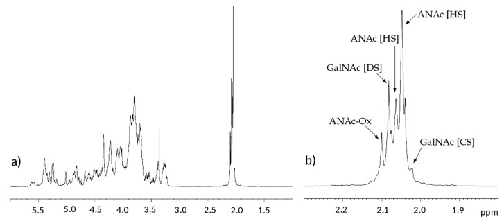
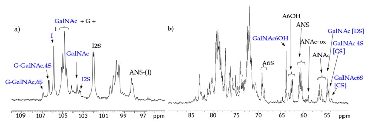
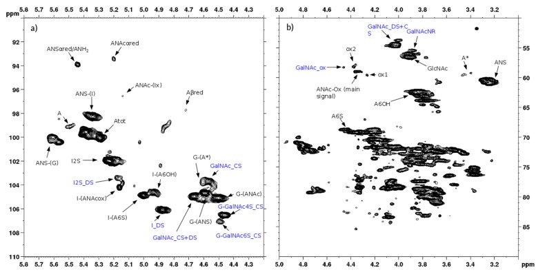
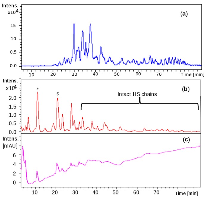
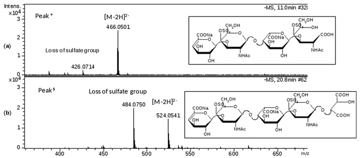
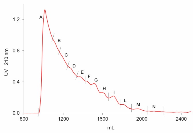
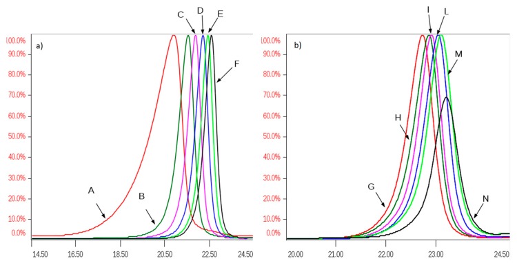
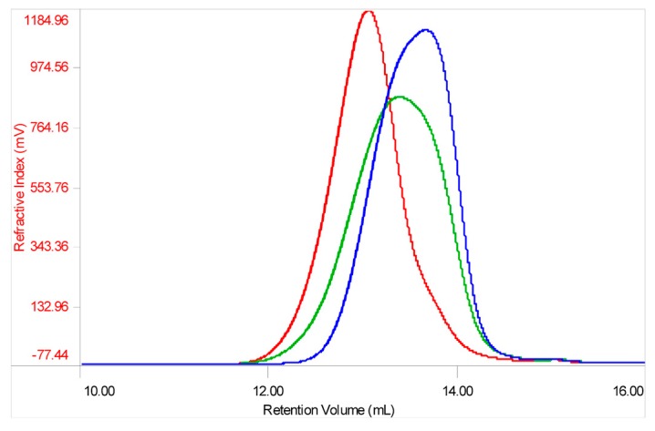
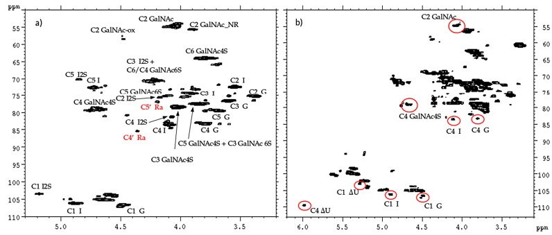
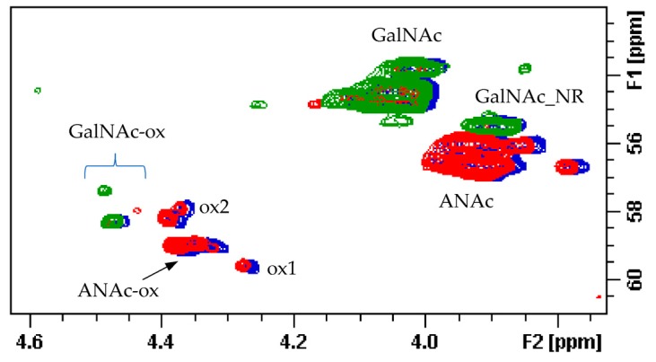
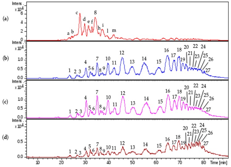
Danaparoid sodium salt, is the active component of ORGARAN, an anticoagulant and antithrombotic drug constituted of three glycosaminoglycans (GAGs) obtained from porcine intestinal mucosa extracts. Heparan sulfate is the major component, dermatan sulfate and chondroitin sulfate being the minor ones. Currently dermatan sulfate and chondroitin sulfate are quantified by UV detection of their unsaturated disaccharides obtained by enzymatic depolymerization. Due to the complexity of danaparoid biopolymers and the presence of shared components, an orthogonal approach has been applied using more advanced tools and methods. To integrate the analytical profile, 2D heteronuclear single quantum coherence (HSQC) NMR spectroscopy was applied and found effective to identify and quantify GAG component signals as well as those of some process signatures of danaparoid active pharmaceutical ingredient (API) batches. Analyses of components of both API samples and size separated fractions proceeded through the determination and distribution of the molecular weight (Mw) by high performance size exclusion chromatographic triple detector array (HP-SEC-TDA), chain mapping by LC/MS, and mono- (¹H and 13C) and bi-dimensional (HSQC) NMR spectroscopy. Finally, large scale chromatographic isolation and depolymerization of each GAG followed by LC/MS and 2D-NMR analysis, allowed the sequences to be defined and components to be evaluated of each GAG including oxidized residues of hexosamines and uronic acids at the reducing ends.

Keywords: component quantitative analysis; danaparoid sodium; low molecular weight glycosaminoglycans; orthogonal multi-analytical methods; sequence and compositional investigations.


Conflict of interest statement

R.v.H and P.d.W. declare that they are employed by Aspen Oss B.V. and that the study was paid for by Aspen Oss B.V.

Figures

Figure 1
Figure 1
Size-exclusion chromatography profile of a danaparoid sample (CAT272): (a) red—refractive index (mV), blue—viscometer (mV) and green—right-angle laser light scattering (mV) detectors; (b) UV (mV) detector.
Figure 2
Figure 2
Example of 1H-NMR spectrum of a danaparoid sample (CAT272): (a) whole spectrum; (b) expansion of the acetyl region.
Figure 3
Figure 3
Example of 13C-NMR spectrum of a danaparoid sample (CAT272): (a) anomeric region; (b) ring carbon region. Black: signals attributed to heparan sulfate (HS); Blue signals attributed to chondroitin sulfate (CS) and dermatan sulfate (DS).
Figure 4
Figure 4
Example of 2D Heteronuclear Single Quantum Coherence (HSQC) NMR of a danaparoid sample (CAT272): (a) anomeric region; (b) ring region. Black: signals attributed to HS; Blue: signals attributed to DS and CS. Abbreviations in Table S1.
Figure 5
Figure 5
Liquid Chromatography-Mass Spectrometry (LC-MS) profiles: (a) one danaparoid sample CAT272; (b) ChABC danaparoid digestion product CAT469; (c) UV chromatogram at 232 nm of CAT469.
Figure 6
Figure 6
Mass spectra of chromatographic peaks * and $, respectively (as labelled in Figure 5b) and corresponding to structures identified by MS/MS fragmentation experiment (data shown in supplementary material Figures S5 and S6): (a) fragment at m/z 466.0501 (z-2; M 934) attributed to ∆U4,2,2(T1); (b) fragment at m/z 524.0541 (z-2; M 1050) attributed to ∆U5,2,2(Ra). The substitution pattern of DS was used.
Figure 7
Figure 7
Preparative size exclusion chromatography (SEC) fractionation chromatographic UV 210 nm profile of a danaparoid (CAT277).
Figure 8
Figure 8
Overlapping of normalized Refractive Index (RI) profiles of SEC fractions of a danaparoid sample (CAT277): (a) fractions A–F, range volume 14–24.5 mL; (b) fractions G–N, range volume 20–24.5 mL.
Figure 9
Figure 9
Structures of oxidized Reducing End (RE) residues compatible with the observed m/z values (the substitution pattern was in accordance with the observed species listed in Table 7).
Figure 10
Figure 10
Mass spectrum displaying the different oxidation forms of oligosaccharide U4,5,0 from M1 to M3 derivative: m/z 552.9907 (z-2, M1 1108) identified as U4,5,0 (T1); m/z 537.9860 (z-2, M2 1078) identified as U4,5,0 (T2); m/z 522.9805 (z-2, M3 1048) identified as U4,5,0 (T3).
Figure 11
Figure 11
Mass signal at m/z 881.086 (z-2): (a) experimental m/z and isotopic distribution; (b) theoretical m/z and isotopic distribution corresponding to the regular structure of U6,6,3 (sum formula C42N3O52H65S6); (c) theoretical m/z and isotopic distribution corresponding to the oxidized structure U6,6,3(T4) (sum formula C41N3O53H61S6).
Figure 12
Figure 12
Example of structures detected in SEC fractions, the modified RE are highlighted in the boxes: HS sequences are displayed in the upper panel with the terminal T1 at the RE and with the uronic acid or the glucosamine at the non reducing end (NRE) in even or odd oligomers, respectively. CS/DS structures are shown in the lower panel (the substitution pattern of DS is used as example because it is more abundant than CS): two possibly reducing ends are T1 and Ra, the galactosamine is placed at the NRE.
Figure 13
Figure 13
Refractive Index profile overlay of a danaparoid sample (CAT272 in green), its enriched CS/DS (red) and HS (blue) fractions.
Figure 14
Figure 14
HSQC NMR spectra of CS/DS (a) and HS (b) fraction of danaparoid CAT272. Signals belonging to CS/DS are circled in red.
Figure 15
Figure 15
Superimposition of HSQC spectra (C2 region) of a danaparoid sample (CAT272 in blue), its enriched HS (red) and CS/DS fractions (green).
Figure 16
Figure 16
LC-MS profiles comparison: (a) danaparoid sample CAT272; (b) CS/DS isolated from CAT271; (c) CS/DS isolated from CAT272; (d) CS/DS isolated from CAT275.
Figure 17
Figure 17
LC-MS profiles comparison: (a) danaparoid sample CAT272; (b) HS isolated from CAT271; (c) HS isolated from CAT272; (d) HS isolated from CAT275.
Figure 18
Figure 18
LC-MS profiles of HS danaparoid (CAT272), nadroparin and heparin digested by heparinases I, II, III.

References

    1. Acostamadiedo J.M., Iyer U.G., Owen J. Danaparoid sodium. Expert Opin. Pharmacother. 2000;1:803–814. doi: 10.1517/14656566.1.4.803. - DOI - PubMed
    1. Nurmohamed M.T., ten Cate H., ten Cate J.W. Low molecular weight heparin(oid)s. Clinical investigations and practical recommendations. Drugs. 1997;53:736–751. doi: 10.2165/00003495-199753050-00002. - DOI - PubMed
    1. Skoutakis V.A. Danaparoid in the prevention of thromboembolic complications. Ann. Pharmacother. 1997;31:876–887. doi: 10.1177/106002809703100715. - DOI - PubMed
    1. Council of Europe. European Pharmacopoeia Commission . European Pharmacopoeia. 6th ed. Strasbourg Council of Europe; Strasbourg, France: 2007. pp. 1644–1646.
    1. Üstün B., Sanders K.B., Dani P., Kellenbach E.R. Quantification of chondroitin sulfate and dermatan sulfate in danaparoid sodium by 1H-NMR spectroscopy and PLS regression. Anal. Bioanal. Chem. 2011;399:629–634. doi: 10.1007/s00216-010-4193-7. - DOI - PubMed