Review
. 2017 Jul 5;10(3):63.
doi: 10.3390/ph10030063.

Nanofiber Scaffolds as Drug Delivery Systems to Bridge Spinal Cord Injury

Affiliations
Review

Nanofiber Scaffolds as Drug Delivery Systems to Bridge Spinal Cord Injury

Angela Faccendini et al. Pharmaceuticals (Basel). .

Abstract

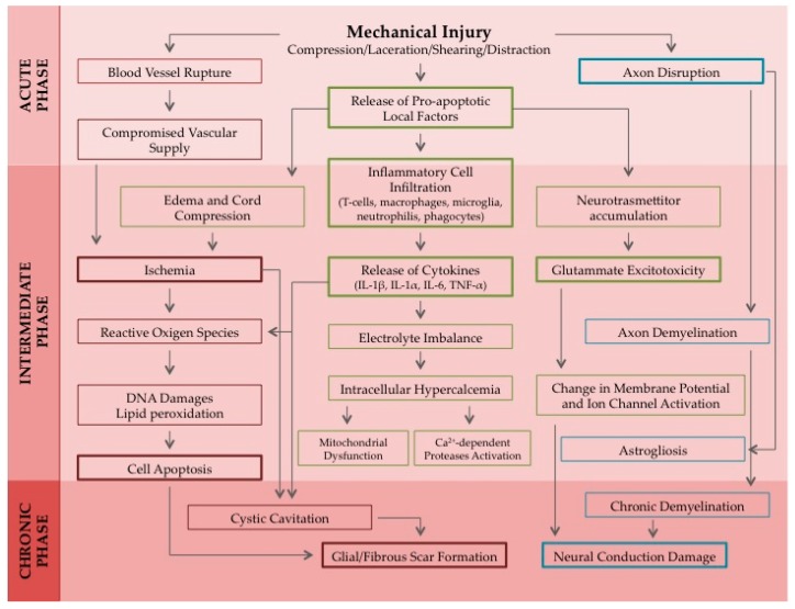
The complex pathophysiology of spinal cord injury (SCI) may explain the current lack of an effective therapeutic approach for the regeneration of damaged neuronal cells and the recovery of motor functions. A primary mechanical injury in the spinal cord triggers a cascade of secondary events, which are involved in SCI instauration and progression. The aim of the present review is to provide an overview of the therapeutic neuro-protective and neuro-regenerative approaches, which involve the use of nanofibers as local drug delivery systems. Drugs released by nanofibers aim at preventing the cascade of secondary damage (neuro-protection), whereas nanofibrous structures are intended to re-establish neuronal connectivity through axonal sprouting (neuro-regeneration) promotion, in order to achieve a rapid functional recovery of spinal cord.

Keywords: electrospinning; nanofibers; neuroprotection; neuroregeneration; spinal cord injury.


Conflict of interest statement

The authors declare no conflict of interest.

Figures

Figure 1
Figure 1
Schematic representation of the pathophysiological response to a spinal cord injury induced by a mechanical trauma. A cascade of vascular, cellular, and biochemical events brings to the progression of the spinal cord damage until the formation of a glial scar. Acronyms: IL-1α, Interleukin 1α; IL-1β, Interleukin 1β; IL-6, Interleukin 6; TNF-α, Tumor Necrosis Factor α.
Figure 2
Figure 2
Nanotechnological approaches for the fabrication of fibrillar structures for the treatment of SCI. (A) Scanning electron micrograph (Zeiss EVO MA10 (Carl Zeiss, Oberkochen, Germany) shows random dextran/alginate fibers; (B) Scanning electron micrograph of carbon nanotubes; scale bars: 250 and 25 μm (inset) (adapted [138]); (C) Scanning electron micrograph of self-assembling nanofibers (adapted from [139]).
Figure 3
Figure 3
Electrospinning process. On the left, the parameters influencing fiber size, morphology, and density are listed; the physicochemical properties of the loaded drugs are also to be considered when the electrospinning technique is used for the fabrication of drug delivery systems. On the right, a schematic representation of the electrospinning apparatus with particular attention on the collector geometry, which is a crucial variable affecting fiber alignment. Scanning electron micrographs (Zeiss EVO MA10 (Carl Zeiss, Oberkochen, Germany)) show random dextran/alginate fibers and aligned polyethilenoxide/alginate ones.

Similar articles

Cited by

References

    1. Kabu S., Gao Y., Kwon B.K., Labhasetwar V. Drug delivery, cell-based therapies, and tissue engineering approaches for spinal cord injury. J. Control. Release. 2015;219:141–154. doi: 10.1016/j.jconrel.2015.08.060. - DOI - PMC - PubMed
    1. Johnson C.D., D’Amato A.R., Gilbert R.J. Electrospun fibers for drug delivery after spinal cord injury and the effects of drug incorporation on fiber properties. Cells Tissues Organs. 2016;202:116–135. doi: 10.1159/000446621. - DOI - PMC - PubMed
    1. Oyinbo C.A. Secondary injury mechanisms in traumatic spinal cord injury: A nugget of this multiply cascade. Acta Neurobiol. Exp. Wars. 2011;71:281–299. - PubMed
    1. Russell C.M., Choo A.M., Tetzlaff W., Chung T.E., Oxland T.R. Maximum principal strain correlates with spinal cord tissue damage in contusion and dislocation injuries in the rat cervical spine. J. Neurotrauma. 2012;29:1574–1585. doi: 10.1089/neu.2011.2225. - DOI - PubMed
    1. Choo A.M., Liu J., Dvorak M., Tetzlaff W., Oxland T.R. Secondary pathology following contusion, dislocation, and distraction spinal cord injuries. Exp. Neurol. 2008;212:490–506. doi: 10.1016/j.expneurol.2008.04.038. - DOI - PubMed

LinkOut - more resources