

Indications for Hip Arthroscopy
- PMID: 28678628
- PMCID: PMC5582699
- DOI: 10.1177/1941738117712675
Indications for Hip Arthroscopy
Abstract
Context: Hip arthroscopy is gaining popularity within the field of orthopaedic surgery. The development and innovation of hip-specific arthroscopic instrumentation and improved techniques has resulted in improved access to the hip joint and ability to treat various hip pathologies.
Evidence acquisition: Electronic databases, including PubMed and MEDLINE, were queried for articles relating to hip arthroscopy indications (1930-2017).
Study design: Clinical review.
Level of evidence: Level 4.
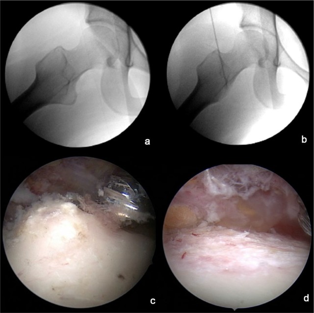
Results: Initially used as a technique for loose body removal, drainage/debridement of septic arthritis, and treatment of pediatric hip disorders, hip arthroscopy is currently used to treat various hip conditions. The recognition of femoroacetabular impingement (FAI) as a source of hip pain in young adults has rapidly expanded hip arthroscopy by applying the principles of osseous correction that were previously described and demonstrated via an open surgical dislocation approach. Hip pathologies can be divided into central compartment, peripheral compartment, peritrochanteric space, and subgluteal space disorders.
Conclusion: Although hip arthroscopy is a minimally invasive procedure that may offer decreased morbidity, diminished risk of neurovascular injury, and shorter recovery periods compared with traditional open exposures to the hip, it is important to understand the appropriate patient selection and indications.
Keywords: femoroacetabular impingement; hip arthroscopy; indications; labral tear; synovial disorders.
Conflict of interest statement
The following authors declared potential conflicts of interest: James R. Ross, MD, is a paid consultant for Smith & Nephew; Christopher M. Larson, MD, is a paid consultant for Smith & Nephew and A3 Surgical and has stock options in A3 Surgical; Asheesh Bedi, MD, is a paid consultant for Arthrex, Inc and has stock/stock options in A3 Surgical.
Figures
References
-
- Adelani MA, Wupperman RM, Holt GE. Benign synovial disorders. J Am Acad Orthop Surg. 2008;16:268-275. - PubMed
-
- Bardakos NV, Villar RN. The ligamentum teres of the adult hip. J Bone Joint Surg Br. 2009;91:8-15. - PubMed
-
- Bedi A, Feeley BT, Williams RJ., 3rd Management of articular cartilage defects of the knee. J Bone Joint Surg Am. 2010;92:994-1009. - PubMed
-
- Bedi A, Ross JR, Kelly BT, Larson CM. Avoiding complications and treating failures of arthroscopic femoroacetabular impingement correction. Instr Course Lect. 2015;64:297-306. - PubMed
-
- Behery O, Siston RA, Harris JD, Flanigan DC. Treatment of cartilage defects of the knee: expanding on the existing algorithm. Clin J Sports Med. 2014;24:21-30. - PubMed
Publication types
MeSH terms
LinkOut - more resources
Full Text Sources
Other Literature Sources
Medical
Research Materials
