. 2017 Jul 13;547(7662):185-190.
doi: 10.1038/nature23002. Epub 2017 Jul 5.

Cryo-EM structures of tau filaments from Alzheimer's disease

Affiliations

Cryo-EM structures of tau filaments from Alzheimer's disease

Anthony W P Fitzpatrick et al. Nature. .

Abstract

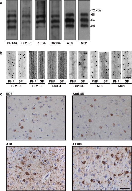
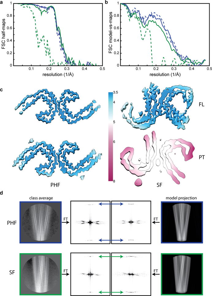
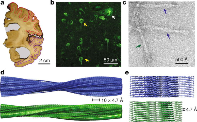
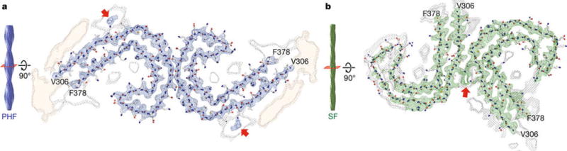
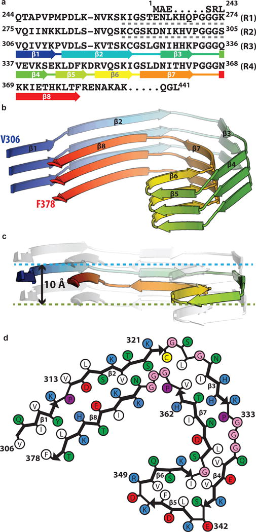
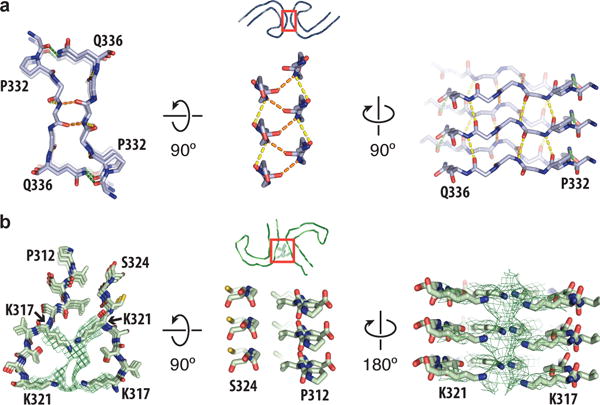
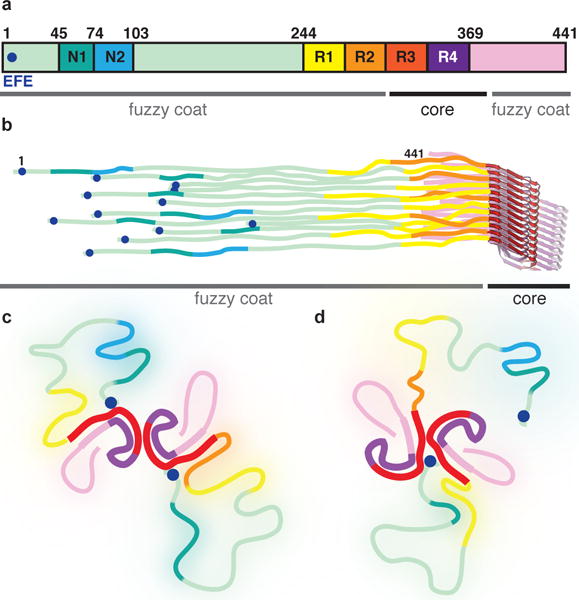
Alzheimer's disease is the most common neurodegenerative disease, and there are no mechanism-based therapies. The disease is defined by the presence of abundant neurofibrillary lesions and neuritic plaques in the cerebral cortex. Neurofibrillary lesions comprise paired helical and straight tau filaments, whereas tau filaments with different morphologies characterize other neurodegenerative diseases. No high-resolution structures of tau filaments are available. Here we present cryo-electron microscopy (cryo-EM) maps at 3.4-3.5 Å resolution and corresponding atomic models of paired helical and straight filaments from the brain of an individual with Alzheimer's disease. Filament cores are made of two identical protofilaments comprising residues 306-378 of tau protein, which adopt a combined cross-β/β-helix structure and define the seed for tau aggregation. Paired helical and straight filaments differ in their inter-protofilament packing, showing that they are ultrastructural polymorphs. These findings demonstrate that cryo-EM allows atomic characterization of amyloid filaments from patient-derived material, and pave the way for investigation of a range of neurodegenerative diseases.


Conflict of interest statement

The authors declare no competing financial interests.

Figures

Extended Data Figure 1
Extended Data Figure 1. Immunolabeling of the brain sample
a,b. Immunolabeling of the sarkosyl-insoluble fraction from the patient’s temporal cortex. Immunoblots (a) using anti-Tau antibodies BR133 (amino-terminus), BR135 (R3), TauC4 (R4), BR134 (carboxy-terminus), AT8 (pS202/pT205) and MC1. Immunogold negative-stain electron microscopy (b) of PHFs and SFs with BR133, BR135, TauC4, BR134, AT8 and MC1. Scale bar, 500 Å. c. Light microscopy of sections from the temporal cortex showing staining of neurofibrillary tangles, neuropil threads and plaque neurites using RD3 (3R), Anti-4R (4R), AT8 and AT100 (pT212/pS214/pT217). Nuclei are counterstained blue. Scale bar, 50 μm.
Extended Data Figure 2
Extended Data Figure 2. PHFs and SFs at various stages in the purification
a–c. Cryo-EM micrographs and reference-free 2D class averages for PHFs (blue insets) and SFs (green insets) for the Tau sample after the sucrose step (a), gel filtration (b), and pronase treatment (c). Examples of PHFs and SFs in the micrographs are indicated with blue and green arrows, respectively. Scale bars, 500 Å. d. Western blots with antibody HT7 (Thermo; catalogue nr. MN1000) of the total lysate, sarkosyl-soluble and sarkosyl-insoluble fractions of HEK 293T cells expressing wild-type 0N4R human tau and treated with (+) or without (−) the sarkosyl-insoluble fraction from the patient’s temporal cortex following gel filtration show that the cryo-EM sample is capable of seeding aggregation of human Tau. e. Densitometric analysis (mean ± standard error of the mean, n=3) of HT7 blots of sarkosyl-insoluble fractions from cells.
Extended Data Figure 3
Extended Data Figure 3. Cryo-EM map and model comparisons
a. Fourier Shell Correlation (FSC) curves between two independently refined half-maps for the FL PHFs (blue, solid); FL SF (green, solid); PT PHF (blue, dashed) and PT SFs (green, dashed). b. FSC curves between the cryo-EM reconstructions and the refined atomic models, using the same colour coding as in a. c. Local resolution estimates for the four cryo-EM reconstructions. d. Comparison of power spectra (the squared amplitudes of the Fourier Transform, FT) of reference-free 2D class averages with those of corresponding projections of the atomic models. In PHFs the approximate 21 screw symmetry between subunits on the two protofilaments leads to off-meridional n=1 Bessel function peaks on the 1/(4.7 Å) layer line (blue arrows). For SFs, where the asymmetric unit consists of two subunits at the same level, one from each protofilament, there is a meridional n=0 Bessel function peak on the 1/(4.7 Å) layer line (green arrows).
Figure 1
Figure 1. Structure of Tau filaments from Alzheimer’s brain
a. Coronal section at the level of the precentral gyrus of the brain used in this study shows an enlarged lateral ventricle (grey background). Grey matter from frontal cortex (red) and temporal cortex (magenta) was used for cryo-EM. b. Thioflavin S staining light microscopy showing abundant neurofibrillary tangles (yellow arrows) and a neuritic plaque (white arrow) in temporal cortex. c. Negatively stained electron micrograph of purified Tau filaments, in which PHFs (blue arrows) and SFs (green arrows) are readily distinguished. d–e. Cryo-EM reconstructions of PHFs (blue) and SFs (green).
Figure 2
Figure 2. Cross-sections of the PHF and SF cryo-EM structures
Cryo-EM density and atomic models of PHFs (a) and SFs (b). Overviews of the helical reconstructions (left) show the orientation of the cross-sectional densities (right). Sharpened, high-resolution maps are shown in blue (PHFs) and green (SFs). Red arrows point at additional densities in contact with K317 and K321. Unsharpened, 4.5 Å low-pass filtered density is shown in grey. Unsharpened density highlighted with an orange background is reminiscent of a less-ordered β-sheet and could accommodate an additional 16 amino acids, which would correspond to a mixture of residues 259–274 (R1) from 3R Tau and residues 290–305 (R2) from 4R Tau.
Figure 3
Figure 3. The common protofilament core
a. Sequence alignment of the four microtubule-binding repeats (R1–R4) with the observed eight β-strand regions coloured from blue to red. The sixteen residues from R1 or R2 that may form an additional, less-ordered β-sheet are indicated with grey dashed lines. b. Rendered view of the secondary structure elements in three successive rungs. c. As in b, but in a view perpendicular to the helical axis, revealing the differences in height along the helical axis within a single molecule. d. Schematic view of the protofilament core.
Figure 4
Figure 4. Protofilament interface in PHFs and SFs
a. Packing between residues 332PGGGQ336 of the two protofilaments in PHFs. Inter-protofilament hydrogen bonds are shown in orange and green. Intra-protofilament hydrogen bonds are shown in yellow. b. Packing between residues 317–324 and residues 312–321 of the protofilaments in SFs. Additional density between the side chains of lysines 317 and 321 is shown as a green mesh.
Figure 5
Figure 5. Schematic representation of full-length Tau filaments
a. Primary structure of the longest Tau isoform in human brain. The two inserts near the amino-terminus are labeled N1 and N2. The microtubule-binding repeats are labeled R1-4. The 7EFE9 motif near the amino-terminus is marked with a blue circle. b. The six different isoforms pack randomly along the helical axis. c. In PHFs, the amino-terminal region of Tau monomers in both protofilaments folds back to form an additional, less ordered β-sheet against β1–β2 of the ordered core, then becomes disordered, and folds back to form an interaction between 7EFE9 and lysines 317 and 321 of the core. d. In SFs, only a single additional β-sheet can form, and the amino-terminus of one of the protofilaments forms part of the protofilament interface.

Comment in

Similar articles

Cited by

References

    1. Wilcock GK, Esiri MM. Plaques, tangles and dementia. A quantitative study. J Neurol Sci. 1982;56:343–356. - PubMed
    1. Ghetti B, et al. Frontotemporal dementia caused by microtubule-associated protein tau gene (MAPT) mutations: a chameleon for neuropathology and neuroimaging. Neuropathol Appl Neurobiol. 2015;41:24–46. - PMC - PubMed
    1. Kidd M. Paired helical filaments in electron microscopy of Alzheimer’s disease. Nature. 1963;197:192–193. - PubMed
    1. Terry RD. The fine structure of neurofibrillary tangles in Alzheimer’s disease. J Neuropathol Exp Neurol. 1963;22:629–642. - PubMed
    1. Yagishita S, Itoh Y, Nan W, Amano N. Reappraisal of the fine structure of Alzheimer’s neurofibrillary tangles. Acta Neuropathol. 1981;54:239–246. - PubMed

Publication types