. 2017 Jul 5;14(7):e1002310.
doi: 10.1371/journal.pmed.1002310. eCollection 2017 Jul.

Ammonium tetrathiomolybdate following ischemia/reperfusion injury: Chemistry, pharmacology, and impact of a new class of sulfide donor in preclinical injury models

Affiliations

Ammonium tetrathiomolybdate following ischemia/reperfusion injury: Chemistry, pharmacology, and impact of a new class of sulfide donor in preclinical injury models

Alex Dyson et al. PLoS Med. .

Abstract

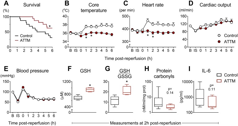
Background: Early revascularization of ischemic organs is key to improving outcomes, yet consequent reperfusion injury may be harmful. Reperfusion injury is largely attributed to excess mitochondrial production of reactive oxygen species (ROS). Sulfide inhibits mitochondria and reduces ROS production. Ammonium tetrathiomolybdate (ATTM), a copper chelator, releases sulfide in a controlled and novel manner, and may offer potential therapeutic utility.

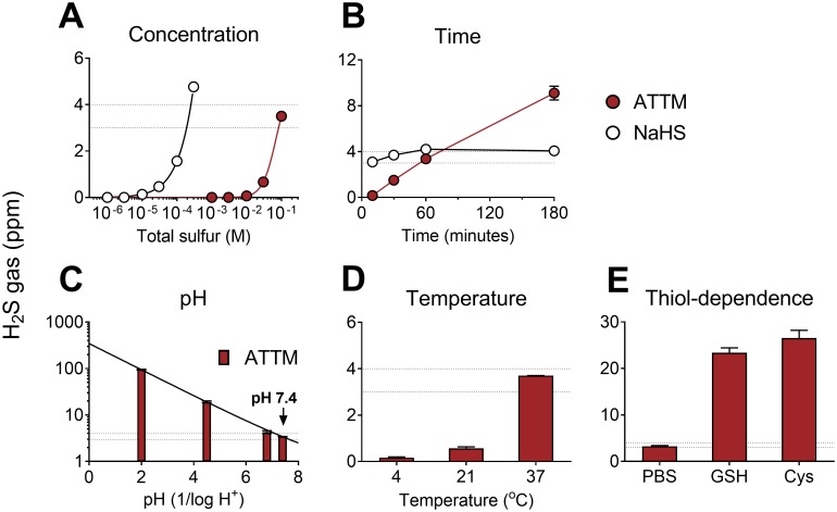
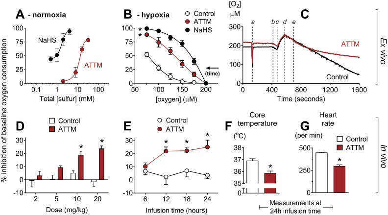
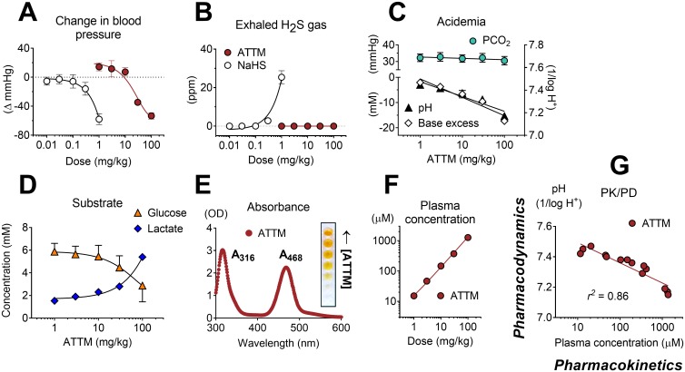
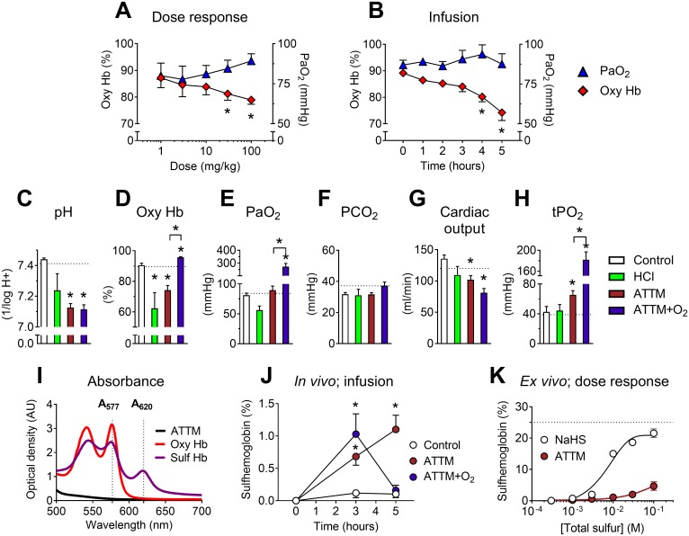
Methods and findings: In vitro, ATTM releases sulfide in a time-, pH-, temperature-, and thiol-dependent manner. Controlled sulfide release from ATTM reduces metabolism (measured as oxygen consumption) both in vivo in awake rats and ex vivo in skeletal muscle tissue, with a superior safety profile compared to standard sulfide generators. Given intravenously at reperfusion/resuscitation to rats, ATTM significantly reduced infarct size following either myocardial or cerebral ischemia, and conferred survival benefit following severe hemorrhage. Mechanistic studies (in vitro anoxia/reoxygenation) demonstrated a mitochondrial site of action (decreased MitoSOX fluorescence), where the majority of damaging ROS is produced.

Conclusions: The inorganic thiometallate ATTM represents a new class of sulfide-releasing drugs. Our findings provide impetus for further investigation of this compound as a novel adjunct therapy for reperfusion injury.


Conflict of interest statement

I have read the journal's policy and the authors of this manuscript have the following competing interests: Magnus Oxygen Ltd are developing ATTM for the treatment of reperfusion injury. AD, JFM and MS are shareholders and MS is head of Magnus Oxygen. AD and ABL are former employees of Magnus Life Science, an historic service platform for Magnus Oxygen. MS is a member of the Editorial Board of PLoS Medicine.

Figures

Fig 1
Fig 1. In vitro release of gaseous H2S from ATTM and NaHS under different environmental conditions.
Comparison of ammonium tetrathiomolybdate (ATTM) and NaHS with changes in (A) concentration and (B) time. In (A), the molarity of each compound was adjusted for equal total sulfur content; drugs were incubated for 1 h at physiological pH (7.4) and temperature (37°C). In (B), fixed concentrations were used: ATTM 100 mM (total sulfur) and NaHS 0.3 mM. The effects of pH, temperature, and the presence of thiols on H2S gas released from ATTM are shown in (C–E). Here, fixed concentrations (100 mM total sulfur) and incubation time (1 h) were employed. Peak H2S concentrations are displayed in parts per million (ppm). The thiols used were reduced glutathione (GSH; 5 mM) and L-cysteine (Cys; 5 mM). The dotted lines reflect typical H2S gas levels (3–4 ppm) obtained from ATTM (100 mM total sulfur) following 1 h incubation at normal physiological pH and temperature. n = 3–6 per group.
Fig 2
Fig 2. Inhibition of oxygen consumption by sulfide-containing drugs.
(A) Ex vivo concentration response curves for ammonium tetrathiomolybdate (ATTM) and NaHS in relative normoxia. Experiments were performed at 150–250 μM O2. In (B), tissues respired to hypoxia. Vehicle, ATTM (0.5 mM, corresponding to 2 mM total sulfur), or NaHS (0.5 mM) was added at 200 μM O2. Note that oxygen consumption in vehicle-treated tissues also decreases at lower [O2] (supply dependence) when tissues respire towards hypoxia. (C) shows a representative trace of tissues respiring to hypoxia, with the timing of the following events indicated: a, sensitivity test; b, addition of tissue to the chamber; c, oxygenation; d, baseline measurements; e, addition of ATTM or vehicle (control). (D) and (E) show the effects of ATTM in vivo following increasing, hourly IV bolus doses or a continuous infusion (10 mg/kg/h), respectively. (F) shows core temperature and (G) shows echocardiography-derived heart rate at the end (24 h) of continuous infusion. Panels A, B, D, and E show percentage inhibition compared to baseline values, before the addition of drugs. *p < 0.05 versus control using a two-way repeated measures ANOVA (plus Bonferroni’s test in D and E) or unpaired t-test (in F and G). n = 3–12 for ex vivo experiments, and n = 4 per group for in vivo studies.
Fig 3
Fig 3. Pharmacokinetic/pharmacodynamic studies.
Maximal changes in mean arterial blood pressure (A) and detection of (peak) exhaled H2S gas (in parts per million [ppm]) (B) following increasing IV bolus doses of ammonium tetrathiomolybdate (ATTM) or NaHS. No exhaled H2S was detectable following ATTM administration. Acid/base interactions following ATTM treatment are shown in (C); the top left y-axis denotes (arterial) partial pressure of carbon dioxide (PCO2); bottom left and right y-axes are (arterial) base excess and pH, respectively. Alterations in (arterial) glucose and lactate following ATTM treatment are shown in (D). (E) shows the absorbance (ultra violet—visible) spectrum of ATTM with (inset) a row of microplate wells used to construct a standard curve. (F) and (G) respectively show changes in ATTM plasma levels (measured using the absorbance peak at 468 nm at 2 min after ATTM administration) against the quantity of drug administered and subsequent (25 min later) changes in arterial pH. n = 3–4/group.
Fig 4
Fig 4. Safety studies.
Effects of ammonium tetrathiomolybdate (ATTM) via either IV bolus dosing (A) or continuous infusion (B) on the arterial partial pressure of oxygen (PaO2) and percentage of oxygenated hemoglobin (oxy Hb). For the continuous infusion study, changes in acid/base balance, hemodynamics, and muscle tissue oxygen tension (tPO2) at experiment end (5 h) are shown in (C–H); dotted lines denote the average baseline value. Where applicable, supplemental oxygen was commenced from 3 h. (I) shows the absorbance spectrum of oxy- and sulfhemoglobin, used for calculation of sulfhemoglobin levels in vivo (in J; infusion study). Note that there was no absorbance overlap between either hemoglobin form and ATTM (1 mM) at λ577/620. Formation of sulfhemoglobin ex vivo using either ATTM or NaHS to spike naïve rat blood is shown in (K). Here, the dotted line represents the maximum sulfhemoglobin level. *p < 0.05 versus baseline (i.e., before the addition of ATTM) in panels A, B, and J using a two-way ANOVA followed by Bonferroni’s testing; *p < 0.05 versus control (and ATTM versus ATTM + O2) in panels C–H using a one-way ANOVA followed by Dunn’s multiple comparison test. n = 5–10 (in vivo), and n = 3 per group (ex vivo) in (K).
Fig 5
Fig 5. Organ-specific ischemia/reperfusion and cellular anoxia/reoxygenation.
Ammonium tetrathiomolybdate (ATTM) confers cardioprotection (A–C, n = 6), neuroprotection (D–F, n = 6), and cytoprotection (G and H, ATTM 5.5 mM, n = 3). In (C), Evans blue dye shows the myocardial area not at risk. In (C) and (F), viable tissue appears pink/red, with non-viable tissue depicted as white/beige. In (G) a representative flow cytometry plot denotes positive (+ve) and negative (−ve) labeling for propidium iodide (PI) and Annexin V, expressed as median fluorescence intensity (MFI). Here, ATTM and controls are depicted in dark red and black, respectively. Live cells (negative for both labels) are shown in the bottom left quadrant and as a percentage of the total in the figure (G, left panel). The dotted line (G, left panel) shows viability in untreated cells (no ischemia/reperfusion or drugs). A representative fluorescence histogram of MitoSOX (mitochondrial superoxide production) is shown in (H), right panel. AAR, area at risk; BNP, B-type natriuretic peptide; IS, infarct size; S100β, S100 calcium binding protein β. *p < 0.05 using an unpaired t-test.
Fig 6
Fig 6. Global ischemia/reperfusion study.
Post-reperfusion survival times are shown in (A) for animals treated with ammonium tetrathiomolybdate (ATTM) versus vehicle (control). *p < 0.05, log-rank test, n = 16 per group. Sequential changes in core temperature, heart rate, cardiac output, and blood pressure are shown (B–E). *p < 0.05 using a two-way repeated measures ANOVA plus Bonferroni test. Note that this test was only performed up to 2 h post-reperfusion due to early mortality. Measurements of (F) blood levels of reduced glutathione (GSH; antioxidant reserve capacity), (G) the ratio of GSH to oxidized glutathione (GSSG) (lower values indicate greater oxidative stress), (H) protein carbonyls (oxidative damage), and (I) interleukin-6 (IL-6; systemic inflammation) were performed at 2 h post-reperfusion, before the onset of significant mortality. *p < 0.05, unpaired t-test.

References

    1. Kalogeris T, Bao Y, Korthuis RJ. Mitochondrial reactive oxygen species: a double edged sword in ischemia/reperfusion vs preconditioning. Redox Biol. 2014;2:702–14. 10.1016/j.redox.2014.05.006 - DOI - PMC - PubMed
    1. GBD 2015 Mortality and Causes of Death Collaborators. Global, regional, and national life expectancy, all-cause mortality, and cause-specific mortality for 249 causes of death, 1980–2015: a systematic analysis for the Global Burden of Disease Study 2015. Lancet. 2016;388:1459–544. 10.1016/S0140-6736(16)31012-1 - DOI - PMC - PubMed
    1. Smith CJ, Denes A, Tyrrell PJ, Di Napoli M. Phase II anti-inflammatory and immune-modulating drugs for acute ischaemic stroke. Expert Opin Investig Drugs. 2015;24:623–43. 10.1517/13543784.2015.1020110 - DOI - PubMed
    1. Rossello X, Yellon DM. A critical review on the translational journey of cardioprotective therapies! Int J Cardiol. 2016;220:176–84. 10.1016/j.ijcard.2016.06.131 - DOI - PubMed
    1. Blackstone E, Morrison M, Roth MB. H2S induces a suspended animation—like state in mice. Science. 2005;308:518 10.1126/science.1108581 - DOI - PubMed