. 2017 Jul 5;13(7):e1006482.
doi: 10.1371/journal.ppat.1006482. eCollection 2017 Jul.

KSHV encoded ORF59 modulates histone arginine methylation of the viral genome to promote viral reactivation

Affiliations

KSHV encoded ORF59 modulates histone arginine methylation of the viral genome to promote viral reactivation

Roxanne C Strahan et al. PLoS Pathog. .

Abstract

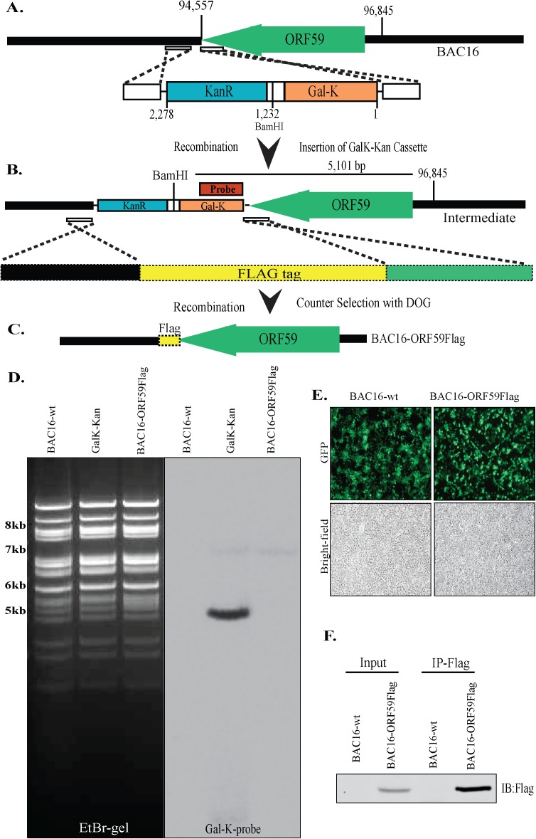
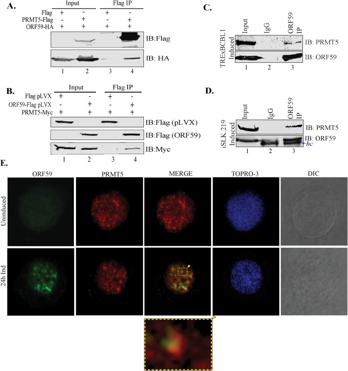
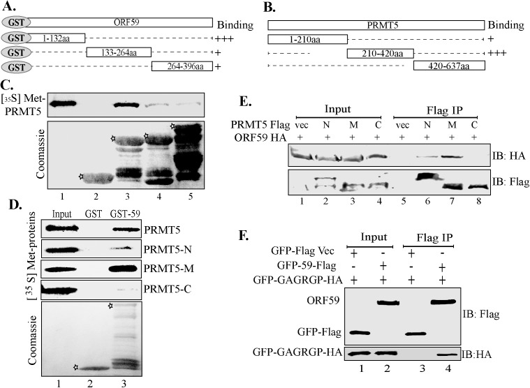
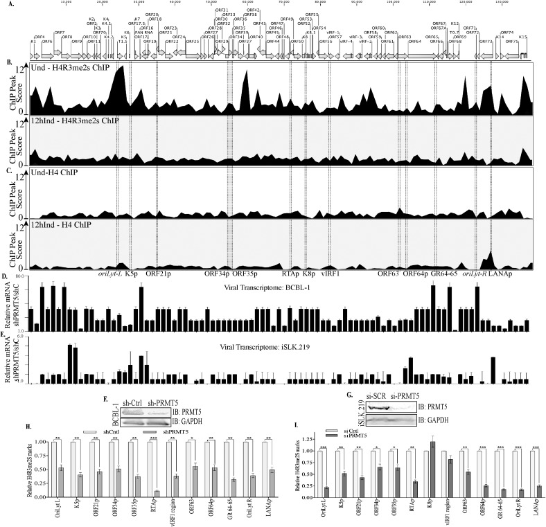
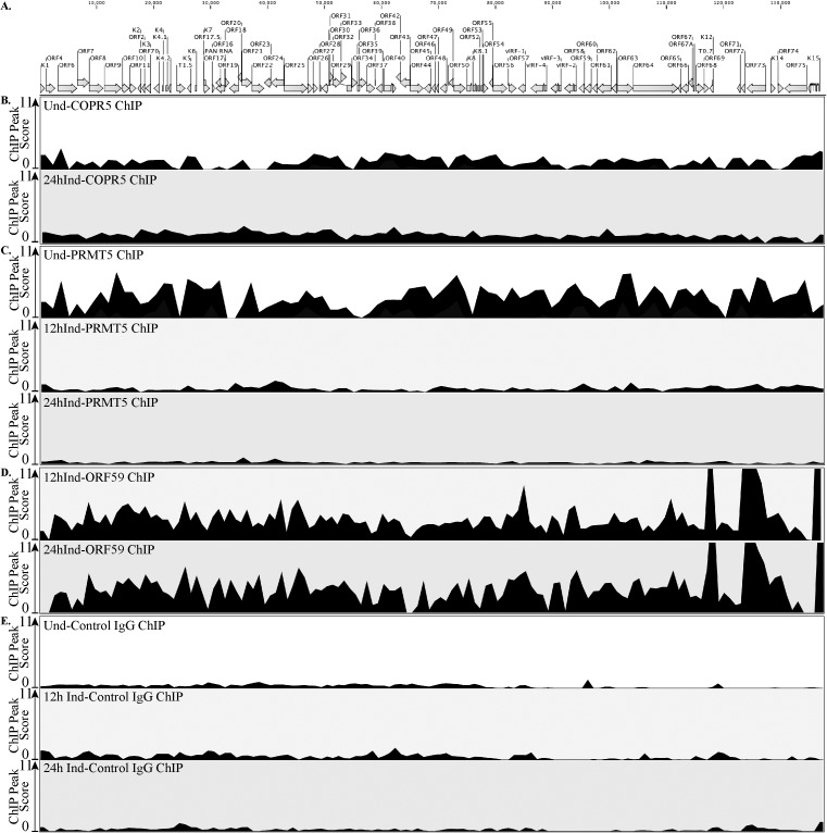
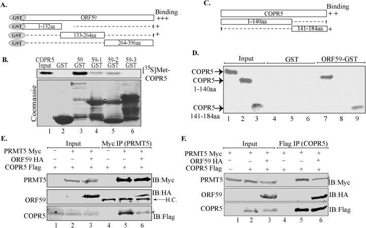
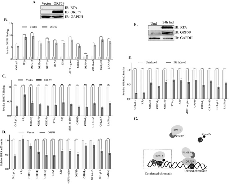
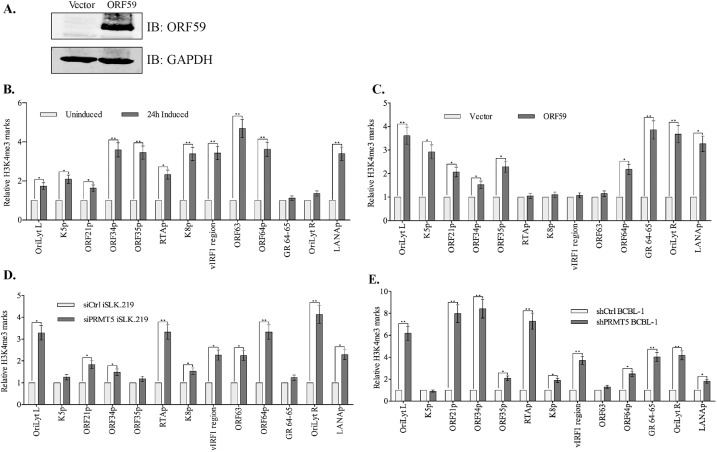
Kaposi's sarcoma associated herpesvirus (KSHV) persists in a highly-ordered chromatin structure inside latently infected cells with the majority of the viral genome having repressive marks. However, upon reactivation the viral chromatin landscape changes into 'open' chromatin through the involvement of lysine demethylases and methyltransferases. Besides methylation of lysine residues of histone H3, arginine methylation of histone H4 plays an important role in controlling the compactness of the chromatin. Symmetric methylation of histone H4 at arginine 3 (H4R3me2s) negatively affects the methylation of histone H3 at lysine 4 (H3K4me3), an active epigenetic mark deposited on the viral chromatin during reactivation. We identified a novel binding partner to KSHV viral DNA processivity factor, ORF59-a protein arginine methyl transferase 5 (PRMT5). PRMT5 is an arginine methyltransferase that dimethylates arginine 3 (R3) of histone H4 in a symmetric manner, one hallmark of condensed chromatin. Our ChIP-seq data of symmetrically methylated H4 arginine 3 showed a significant decrease in H4R3me2s on the viral genome of reactivated cells as compared to the latent cells. Reduction in arginine methylation correlated with the binding of ORF59 on the viral chromatin and disruption of PRMT5 from its adapter protein, COPR5 (cooperator of PRMT5). Binding of PRMT5 through COPR5 is important for symmetric methylation of H4R3 and the expression of ORF59 competitively reduces the association of PRMT5 with COPR5, leading to a reduction in PRMT5 mediated arginine methylation. This ultimately resulted in a reduced level of symmetrically methylated H4R3 and increased levels of H3K4me3 marks, contributing to the formation of an open chromatin for transcription and DNA replication. Depletion of PRMT5 levels led to a decrease in symmetric methylation and increase in viral gene transcription confirming the role of PRMT5 in viral reactivation. In conclusion, ORF59 modulates histone-modifying enzymes to alter the chromatin structure during lytic reactivation.


Conflict of interest statement

The authors have declared that no competing interests exist.

Figures

Fig 1
Fig 1. Schematic illustrating the flag epitope tagged ORF59 BACmid (BAC16-ORF59Flag) generation via homologous recombination.
A-C. The flag epitope tag was inserted at the C-terminus of ORF59 by recombineering and GalK-KanR counter selection. Presence of GalK-Kan cassette was confirmed by BamHI digestion and Southern hybridization with GalK probe indicated in red (probe). Sequencing confirmed the insertion of Flag epitope tag. D. EtBr gel of BamHI digested BAC16-wt (lane 1), Intermediate containing GalK-KanR cassette (lane 2) and BAC16-ORF59Flag (lane 3). Southern blot with GalK probe showed expected 5,101bp band in the intermediate (lane 2). E. The HEK293L cells harbored stable BAC16-wt and BAC16-ORF59Flag. Green panels show the expression of GFP confirming the presence of Bac16WT or Bac16-ORF59Flag and the gray panels are the corresponding bright-field images. F. Flag tagged ORF59 was confirmed in the cell lysate (input lane) and by α–Flag immunoprecipitation and western blot (IP-Flag lane).
Fig 2
Fig 2. ORF59 associates with PRMT5.
A. Binding between full-length ORF59 and PRMT5 was established by cotransfecting ORF59-HA and PRMT5-Flag plasmids and immunoprecipitating with anti-Flag antibody. The western blot shows specific binding between PRMT5 and ORF59 (lane 4), as the vector alone did not precipitate any ORF59 (lane 3). B. A reverse co-immunoprecipitation demonstrates binding between ORF59 and PRMT5. ORF59-Flag was cotransfected with PRMT5-Myc and the lysate was subjected to immunoprecipitation with anti-Flag antibody. These two proteins showed specific association as lane 4 shows PRMT5-Myc band but the vector alone did not precipitate any PRMT5-Myc. C, and D. KSHV-positive. TRExBCBL1-Rta cells or iSLK.219 cells were induced with doxycycline/NaB before harvesting for immunoprecipitation of ORF59. Lane 3 demonstrates a specific association between ORF59 and endogeneous PRMT5 (lane 3) while IgG control did not show detectable levels of PRMT5 (lane 2). Heavy chain-hc. E. Uninduced or 24h doxycycline induced TRExBCBL1-Rta cells were fixed on glass coverslips and stained for ORF59 and PRMT5 localization using specific antibodies. These cells were counter-stained with TO-PRO3 for the detection of nuclei. Images of showed colocalization of ORF59 and PRMT5 in the cell nucleus. Merge signals were magnified to demonstrate colocalization. iSLK.219 cells were not included in the IFA assay because of GFP backbone in the viral genome and express RFP after induction.
Fig 3
Fig 3. ORF59 binds to the middle (210-420aa) segment of PRMT5.
A. Schematic of ORF59 and its truncations fused to GST. B. Domains of PRMT5 used in binding assays. C. In vitro translated 35S-methionine labeled-PRMT5 subjected to binding with truncated domains of ORF59 fused to GST. The N-terminal domain, 1-132aa of ORF59 bound most strongly with PRMT5 (lane 3), while control GST (lane 2) did not show any detectable band demonstrating specificity. Asterisks on the coomassie gel indicate GST fused segments of ORF59 used in the binding assay. D. In vitro translated 35S-methionine labeled PRMT5 full length and its truncation (amino-N, middle-M and carboxyl-C) mutants were subjected to binding with control GST (GST) (lane 2) or GST-59 (lane 3). PRMT5 residues 210-420aa (middle segment) associated most strongly with GST-ORF59 (lane 3). Asterisks indicate Control GST (lane 2) and full-length ORF59 fused GST (lane 3) in the coomassie image. E. Flag-tagged PRMT5 truncations (amino-N, middle-M and carboxyl-C) mutants were co-expressed with HA-epitope tagged ORF59 and lysates were immunoprecipitated with anti-Flag antibody. PRMT5 210-420aa immunoprecipitated ORF59 specifically as compared to Flag-vector (compare lanes 5 with 7) and PRMT5 1-210aa also showed a weaker association with ORF59 (lane 6). F. The middle domain of PRMT5 containing the catalytic domain (GAGRGP) was fused to GFP and HA and co-expressed with ORF59-Flag or vector control GFP-Flag and lysates were immunoprecipitated with anti-Flag antibody. ORF59 associated with the GFP-HA-tagged PRMT5 catalytic domain but vector control GFP alone did not (compare lane 4 with 3, IB: HA panel).
Fig 4
Fig 4. Symmetric di-methylation of H4R3 (H4R3me2s) levels were significantly reduced in lytic reactivated and PRMT5 depleted cells.
A. Schematic of the KSHV genome with open reading frames indicated. B. Chromatin from uninduced TRExBCBL1-RTA cells was immunoprecipitated with anti-H4R3me2s antibody followed by next-generation sequencing of the ChIP DNA (ChIP-Seq) and detection of ChIP peaks. Chromatin from 12h doxycycline-induced TRExBCBL1-RTA cells was immunoprecipitated with anti-H4R3me2s antibody followed by next-generation sequencing of the ChIP DNA (ChIP-Seq) and detection of ChIP peaks. The peak score represents peak height. The prevalence of H4R3me2s marks was reduced on viral genome undergoing lytic reactivation. C. Histone H4 on the latent and 12h induced TRExBCBL1-RTA cells were determined by mapping the reads to the KSHV reference genome and then detecting ChIP peaks. D. Relative expression of viral genes in PRMT5 depleted (shPRMT5) TRExBCBL1-Rta cells as compared to the control (shCtrl) cells. Viral genes detected by qPCR showed an enhanced gene transcription in the absence of PRMT5 relative to the control. E. Relative expression of viral genes in PRMT5 depleted (siPRMT5) iSLK.219 cells as compared to the control, scrambled (si-Ctrl) cells. Transcriptome analysis of entire KSHV genome by qPCR showed an enhanced expression of many genes in PRMT5 depleted (si-PRMT5) as compared to the control (si-Ctrl) cells. F. BCBL-1 cells stably transduced with PRMT5 shRNA expressing lentiviruses showed depletion of PRMT5 levels as compared to the control shRNA transduced cells. G. iSLK.219 cells were transfected with si-Control or si-PRMT5 showed reduction of PRMT5 in si-PRMT5 cells. H. BCBL-1 cells depleted with PRMT5 in panel F were subjected for chromatin immunoprecipitation with anti-H4R3me2s antibody and analyzed by qPCR at selected region at promoters, which displayed differential levels of H4R3me2s in our ChIP-Seq results. PRMT5-depletion displayed significantly lower levels of H4R3me2s than shControl cells al all the tested regions. I. iSLK.219 cells depleted with PRMT5 from panel G were subjected ChIP assay with anti-H4R3me2s antibody and analyzed by qPCR at the same regions used on panel H showed almost similar reduction of H4R3me2s bound chromatin at those regions.
Fig 5
Fig 5. ORF59 binding to the viral genome during reactivation reduced the levels of PRMT5 bound to the viral chromatin.
A. Schematic of the KSHV genome with ORFs. B-E. Uninduced, 12h Ind or 24h Ind (as indicated) TRExBCBL1-RTA cells were harvested, fixed, chromatin was sheared and then precipitated using specific anti-COPR5, anti-PRMT5, anti-ORF59, or anti-IgG antibodies. ChIP DNA was isolated and used to prepare sequencing libraries which were analyzed on the Illumina NextSeq500 instrument. Reads were mapped to the KSHV genome and significant ChIP peaks were identified using “ChIP Seq” tool of CLC Genomics Workbench and the ChIP peaks are presented as peak score. B. COPR5 binding on viral chromatin during uninduced (Und) and 24h induced (24h Ind) cells. C. PRMT5 binding on viral chromatin during uninduced (Und), 12 and 24h induced (12h Ind, 24h Ind) cells. D. ORF59 binding on viral chromatin during 12 and 24h induced (12h Ind, 24h Ind) cells. E. Control-IgG ChIP was performed on latent, 12h induced, and 24h induced cells and the reads were mapped to the KSHV genome and analyzed by ChIP-peak calling software to show protein binding specificity.
Fig 6
Fig 6. ORF59 competitively disrupts PRMT5’s binding with its binding ligand, COPR5.
A. Schematic representation of GST-fused ORF59 and its truncation mutants. B. In vitro translated 35S-methionine labeled COPR5 was subjected to a binding assay with ORF59-GST and its truncation mutants, 59–1 (1-132aa), 59–2 (133aa-264aa) and 59–3 (264-396aa). Full-length ORF59 bound most strongly to COPR5 although ORF59-1 and ORF59-2 also associated with COPR5 with lower affinity (compare lane 3 with lane 4 and 5). Asterisks in the coomassie panel indicate control GST (lane 2) GST-fused ORF59 (lane 3) and its truncations (lanes 4–6). C. Schematic representation of COPR5 with its histone binding truncation (1-140aa), and PRMT5 binding truncation (141-184aa). D. In vitro translated 35S-methionine labeled COPR5 and its truncations were subjected to binding with control GST (lanes 4–6) or GST fused ORF59 (lanes 7–9). Lanes 1–3 show input levels of in vitro translated COPR5 segments. Full length COPR5 and the PRMT5 binding domain of COPR5 (141-184aa) bound to the GST fused ORF59 but not the control GST (compare lanes 7 and 9 to 4 and 6). E. PRMT5-Myc, ORF59 HA, and COPR5-Flag were cotransfected and the lysates were immunoprecipitated with anti-Myc antibody to precipitate PRMT5, which precipitated COPR5, as expected (lane 5). However, in the cells expressing ORF59 the association between PMRT5 and COPR5 was reduced, lane 6. ORF59 immunoprecipiated from the complex confirming its association with PRMT5 (lane 6, IB:HA panel). F. Competitive binding was also performed by immoprecipitating COPR5 in presence of of ORF59 (lanes 3). The level of COPR5 bound PRMT5 was reduced (compare lanes 5 and 6).
Fig 7
Fig 7. ORF59 orchestrates chromatin modification independent of RTA.
A. iSLK-BAC16-RTASTOP cells were transfected with ORF59 or control vector and the expression was confirmed by WB. B. Chromatin from the iSLK-BAC16-RTASTOP with ORF59 or vector transfected cells were immunoprecipitated with anti-ORF59 antibody and analyzed for its binding at various viral promoters. ORF59 overexpression in these RTA-depleted cells resulted in specific binding of ORF59 to the viral promoter targets when compared to the cells without ORF59 expression. C. ChIP with PRMT5 was performed to determine the binding of PRMT5 in presence of ORF59 at those selected regions of the viral genome, which showed reduced binding, as detected in the lytically reactivated cells. D. ChIP assay with anti-H4R3me2s antibody was performed to determine the levels of chromatin bound to symmetrically methylated H4R3. In alliance with PRMT5 being depleted, the levels of H4R3me2s are reduced at the viral promoters in the presence of overexpressed ORF59. E. iSLK-BAC16-RTASTOP cells were either un-induced, or lytically reactivated by doxycycline for 24h, expressed RTA and ORF59. F. These doxycycline induced or uninduced iSLK-BAC16-RTASTOP cells were subjected for ChIP with anti-H4R3me2s antibody for detecting chromatin bound to H4R3me2s chromatin. Consistent with our previous results, H4R3me2s bound chromatin was less prevalent at viral promoters during induction as compared to the uninduced, latent cells. G. Proposed model of the mechanism by which ORF59 depletes PRMT5 binding from the chromatin to facilitate the relaxation of viral chromatin and gene transcription.
Fig 8
Fig 8. Loss of H4R3me2s corresponds with an enrichment of H3K4me3 on viral chromatin.
A. iSLK-BAC16-RTASTOP cells were transfected with ORF59 or control vector plasmids to express only ORF59 in these latent cells. B. iSLK-BAC16-RTASTOP were induced with doxycycline for 24h, fixed, and chromatins were sheared and immunoprecipitated with anti-H3K4me3 antibody. Congruent with previous reports, the levels H3K4me3 bound chromatin were enriched early during lytic reactivation at representative regions. C. iSLK-BAC16-RTASTOP cells expressing ORF59 were subjected for ChIP with anti-H3K4me3 antibody, which showed an enrichment of H3K4me3 at various viral promoters calculated relative to the vector transfected cells. D. iSLK.219 cells transiently transfected with control or PRMT5 siRNA to deplete PRMT5 were subjected for ChIP with anti-H3K4me3 antibody. Relative amounts of H3K4me3 bound chromatin was enhanced ion In PRMT5 depleted cells as compared to the control cells. E. Similarly, BCBL-1 cells depleted for PRMT5 by stable transducing specific shRNA lentiviral vector showed an enhanced level of H3K4me3 bound chromatin as compared to control sh RNA lentiviral transduced cells shown at representative regions.
Fig 9
Fig 9. Expression of ORF59 triggers expression of viral genes.
293LBac36WT or ORF59 deleted (293LBac36Δ59) cells were transfected with RTA to induce lytic reactivation. Cells were harvested 48h post transfection cells for the detection of viral mRNA in a real-time qPCR assay. Expression of many viral genes were reduced in ORF59 deleted cells and the genes highlighted in green are immediate early, blue-early genes and red are the late genes.

Similar articles

Cited by

References

    1. Cesarman E, Chang Y, Moore PS, Said JW, Knowles DM. Kaposi's sarcoma-associated herpesvirus-like DNA sequences in AIDS-related body-cavity-based lymphomas. N Engl J Med. 1995;332(18):1186–91. doi: 10.1056/NEJM199505043321802 . - DOI - PubMed
    1. Chang Y, Cesarman E, Pessin MS, Lee F, Culpepper J, Knowles DM, et al. Identification of herpesvirus-like DNA sequences in AIDS-associated Kaposi's sarcoma. Science. 1994;266(5192):1865–9. Epub 1994/12/16. . - PubMed
    1. Soulier J, Grollet L, Oksenhendler E, Cacoub P, Cazals-Hatem D, Babinet P, et al. Kaposi's sarcoma-associated herpesvirus-like DNA sequences in multicentric Castleman's disease. Blood. 1995;86(4):1276–80. . - PubMed
    1. Uldrick TS, Wang V, O'Mahony D, Aleman K, Wyvill KM, Marshall V, et al. An Interleukin-6-Related Systemic Inflammatory Syndrome in Patients Co-Infected with Kaposi Sarcoma-Associated Herpesvirus and HIV but without Multicentric Castleman Disease. Clin Infect Dis. 2010;51(3):350–8. Epub 2010/06/30. doi: 10.1086/654798 . - DOI - PMC - PubMed
    1. Dittmer D, Lagunoff M, Renne R, Staskus K, Haase A, Ganem D. A cluster of latently expressed genes in Kaposi's sarcoma-associated herpesvirus. J Virol. 1998;72(10):8309–15. ; PubMed Central PMCID: PMCPMC110196. - PMC - PubMed