. 2017 Jul 6;15(1):58.
doi: 10.1186/s12915-017-0396-0.

Q&A: using Patch-seq to profile single cells

Affiliations

Q&A: using Patch-seq to profile single cells

Cathryn R Cadwell et al. BMC Biol. .

Abstract

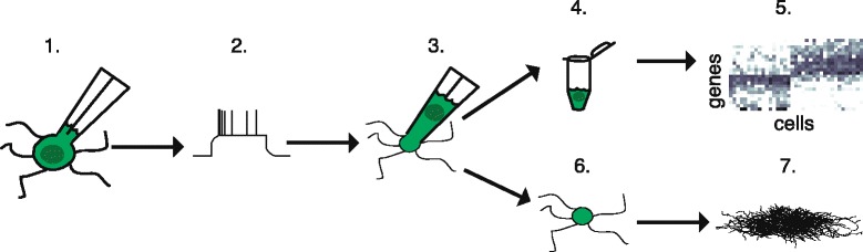
Individual neurons vary widely in terms of their gene expression, morphology, and electrophysiological properties. While many techniques exist to study single-cell variability along one or two of these dimensions, very few techniques can assess all three features for a single cell. We recently developed Patch-seq, which combines whole-cell patch clamp recording with single-cell RNA-sequencing and immunohistochemistry to comprehensively profile the transcriptomic, morphologic, and physiologic features of individual neurons. Patch-seq can be broadly applied to characterize cell types in complex tissues such as the nervous system, and to study the transcriptional signatures underlying the multidimensional phenotypes of single cells.


Figures

Fig. 1.
Fig. 1.
Overview of Patch-seq technique. Access to the intracellular compartment of a single neuron is gained by whole-cell patch clamp (step 1) and the electrical properties of the cell, such as its firing pattern in response to depolarizing current injection, are recorded (step 2). The intracellular contents are aspirated into the patch pipette (step 3) and collected in a PCR tube (step 4) for downstream RNA-sequencing (step 5). The tissue slice, which retains the collapsed cell body and fine processes of the cell (step 6), is subjected to immunohistochemical staining to visualize the complex morphology of the cell (step 7). Adapted by permission from Macmillan Publishers Ltd: Nature Biotechnology [1], copyright (2016)
Fig. 2.
Fig. 2.
Combined Patch-seq and morphological recovery. Immunohistochemical staining (left, scale bar 50 μm) and full-length amplified cDNA Bioanalyzer profile (right) from a layer 2/3 pyramidal neuron. ERCC spike-in RNA was used as a positive control in this experiment and gives rise to the distinct peaks at ~600 bp and ~1100 bp in the Bioanalyzer profile
Fig. 3.
Fig. 3.
Collapse of cell body during aspiration into patch pipette. Two example neurons patched in vivo under two-photon guidance using a green fluorescent dye in the patch pipette. In both cases, the cell body was noted to decrease in size dramatically after aspirating the cell contents into the pipette. Adapted by permission from Macmillan Publishers Ltd: Nature Biotechnology [1], copyright (2016)

References

    1. Cadwell CR, Palasantza A, Jiang X, Berens P, Deng Q, Yilmaz M, et al. Electrophysiological, transcriptomic and morphologic profiling of single neurons using Patch-seq. Nat Biotechnol. 2016;34:199–203. doi: 10.1038/nbt.3445. - DOI - PMC - PubMed
    1. Fuzik J, Zeisel A, Mate Z, Calvigioni D, Yanagawa Y, Szabo G, et al. Integration of electrophysiological recordings with single-cell RNA-seq data identifies neuronal subtypes. Nat Biotechnol. 2016;34:175–83. doi: 10.1038/nbt.3443. - DOI - PMC - PubMed
    1. Zeisel A, Munoz-Manchado AB, Codeluppi S, Lonnerberg P, La Manno G, Jureus A, et al. Brain structure. Cell types in the mouse cortex and hippocampus revealed by single-cell RNA-seq. Science. 2015;347:1138–42. doi: 10.1126/science.aaa1934. - DOI - PubMed
    1. Tasic B, Menon V, Nguyen TN, Kim TK, Jarsky T, Yao Z, et al. Adult mouse cortical cell taxonomy revealed by single cell transcriptomics. Nat Neurosci. 2016;19:335–46. doi: 10.1038/nn.4216. - DOI - PMC - PubMed
    1. Macosko EZ, Basu A, Satija R, Nemesh J, Shekhar K, Goldman M, et al. Highly parallel genome-wide expression profiling of individual cells using nanoliter droplets. Cell. 2015;161:1202–14. doi: 10.1016/j.cell.2015.05.002. - DOI - PMC - PubMed

Publication types