. 2017 Jul 18;114(29):7689-7694.
doi: 10.1073/pnas.1707741114. Epub 2017 Jul 5.

Identification of pathogenic gene mutations in LMNA and MYBPC3 that alter RNA splicing

Affiliations

Identification of pathogenic gene mutations in LMNA and MYBPC3 that alter RNA splicing

Kaoru Ito et al. Proc Natl Acad Sci U S A. .

Abstract

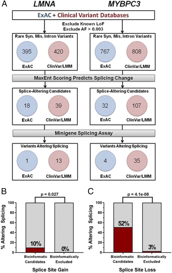
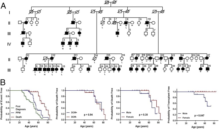
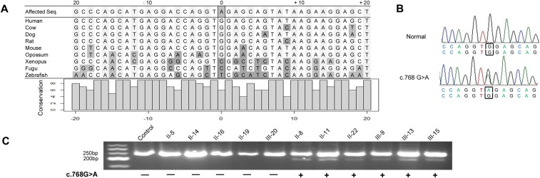
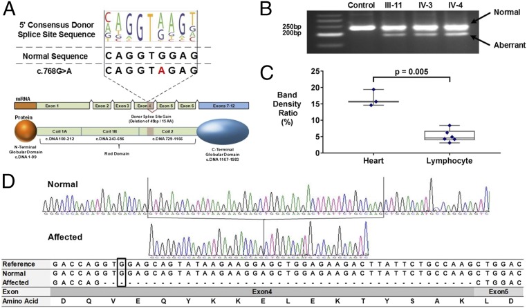
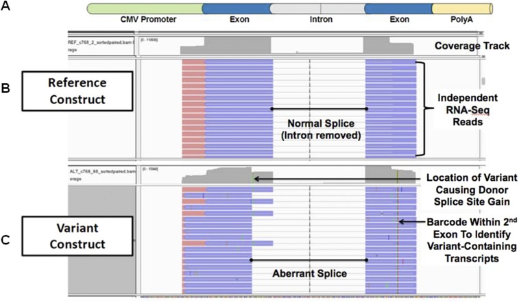
Genetic variants that cause haploinsufficiency account for many autosomal dominant (AD) disorders. Gene-based diagnosis classifies variants that alter canonical splice signals as pathogenic, but due to imperfect understanding of RNA splice signals other variants that may create or eliminate splice sites are often clinically classified as variants of unknown significance (VUS). To improve recognition of pathogenic splice-altering variants in AD disorders, we used computational tools to prioritize VUS and developed a cell-based minigene splicing assay to confirm aberrant splicing. Using this two-step procedure we evaluated all rare variants in two AD cardiomyopathy genes, lamin A/C (LMNA) and myosin binding protein C (MYBPC3). We demonstrate that 13 LMNA and 35 MYBPC3 variants identified in cardiomyopathy patients alter RNA splicing, representing a 50% increase in the numbers of established damaging splice variants in these genes. Over half of these variants are annotated as VUS by clinical diagnostic laboratories. Familial analyses of one variant, a synonymous LMNA VUS, demonstrated segregation with cardiomyopathy affection status and altered cardiac LMNA splicing. Application of this strategy should improve diagnostic accuracy and variant classification in other haploinsufficient AD disorders.

Keywords: LMNA; MYBPC3; VUS; cardiomyopathy; splicing.


Conflict of interest statement

Conflict of interest statement: C.E.S. and J.G.S. are founders of and own shares in Myokardia Inc., a startup company that is developing therapeutics that target the sarcomere.

Figures

Fig. 1.
Fig. 1.
Schematics of gene splice signals and a functional assay of potential splice-altering variants. (A) A gene segment containing two exons (blue) and flanking introns (gray) with consensus 9-bp splice donor and 23-bp splice acceptor sequences is shown. The nucleotide letter sizes (drawn using Pictogram; genes.mit.edu/pictogram.html) denote use of that base in splicing across the genome. Dinucleotides GT (donor site) and AG (acceptor site) are invariant. Normally spliced transcript (red) that excludes intron sequence is depicted. (B) Functional assay of splicing candidates used a 1,200-bp minigene construct comprised of CMV promoter, exon–intron–exon sequence, a 2-bp barcode to identify the construct, and SV40polyA sequence. Exon–intron–exon test sequences were encoded on a synthetic 500-bp oligonucleotide that limited the inclusion of intron sequences to 200 bp (further details provided in Materials and Methods and Datasets S3 and S4). A normally spliced minigene-derived transcript (red) that excludes intron sequence is depicted. (C) Splice-altering variants detected by functional assays. Variants that destroy donor or acceptor signals prevent normal splicing (“x”) and yield transcripts with included introns, partial deletions, or entirely skipped exons. [Note that analyses reported excluded all variants that altered the invariant GT (donor site) and AG (acceptor site) sequences.] Variants that create a novel splice signal (donor or acceptor gain, “”) inappropriately delete sequences from the 5′ or 3′ exon. Novel splice sites that occur within introns and insert sequences are not depicted.
Fig. S1.
Fig. S1.
Thresholds for selection of candidate splice altering variants (Materials and Methods). Youden’s index (index = sensitivity + specificity − 1) was maximized to set a MaxEnt score value below which variants were not predicted to lead to 5′ donor or 3′ acceptor splice site gain. (A) At the 5′ donor splice site, Youden’s index was maximized when MaxEnt score = 4.1. (B) At the 3′ acceptor splice site, Youden’s index was maximized when MaxEnt score = 4.4. (C) A decision tree depicts the algorithm used for selection of candidate splice-altering variants. Variants predicted to cause loss of a donor or acceptor splice site (contained within splice site, ΔMaxEnt < 0) were selected for cell splicing assays. Variants predicted to create a gain of donor splice site (outside of splice site, ΔMaxEnt > 0) and had MaxEntvar score > 4.1 were selected for the minigene splicing assay. Similarly, variants predicted to create a gain of acceptor splice site (outside of splice site, ΔMaxEnt > 0) and had MaxEntvar score > 4.4 were selected for the minigene assay.
Fig. 2.
Fig. 2.
Prioritization and functional assessment of synonymous, missense, or intron variants in LMNA and MYBPC3. (A) Rare (allele frequency <0.003) variants in LMNA (n = 815) and MYBPC3 (n = 1,575) variants reported in the ExAC and clinical diagnostic databases were assessed by computational algorithms to prioritize LMNA (n = 57) and MYBPC3 (n = 139) variants for functional assays. Fourteen LMNA and 39 MYBPC3 variants altered splicing in the cell-based assay. (B) Percent of variants successfully conferring splice site gain among bioinformatically prioritized candidates and bioinformatically excluded variants. A total of 12 out of 117 variants predicted to alter splicing did so in the minigene assay. Of 39 variants tested with MaxEnt scores below the threshold set for creation a new splice site, zero altered splicing (P = 0.03). (C) Percent of variants leading to splice site loss among bioinformatically prioritized candidates and bioinformatically excluded variants. A total of 41 out of 79 variants predicted to lead to splice site loss did so in the minigene assay. Of 35 variants predicted to have no effect on the splice site, one was found to abrogate normal splicing (P = 4e-08).
Fig. S2.
Fig. S2.
The workflow of the entire minigene splicing assay. All scripts are available online at https://github.com/SplicingVariant/SplicingVariants_Beta. (A) Variants are extracted from the clinical diagnostic and ExAC databases. Variants were initially screened to exclude all variants with allele frequencies >0.003, as well as variants that led to nonsense, frameshift, or altered the canonical GT or AG bases in consensus splice sites. (B) Variant positions in a given transcript were retrieved from Biomart database and MaxEnt scores for both reference allele and variant allele were calculated. The decision tree to select candidate splice altering variants based on MaxEnt scoring is depicted in Fig. S1C. Candidate variants were used to design mutant and reference constructs as described in Materials and Methods. (C) Reference and variant minigene constructs are assembled using a combination of synthetic oligonucleotides and PCR. Minigene constructs are pooled and transfected into HEK293 cells. After 24 h, RNA is extracted, converted into a cDNA library, and sequenced. (D) The sequence data are demultiplexed and analyzed for quantitative assessment of no splice, normal splice, and aberrant splicing. The status of splicing is determined comparing statistics between reference- and variant-containing minigenes.
Fig. 3.
Fig. 3.
A rare synonymous LMNA variant segregates with DCM and conduction system disease. (A) The four-generation pedigree demonstrated autosomal dominant inheritance of the LMNA c.768 G > A variant (LOD = 20.02, θ = 0) and cardiomyopathy. (Solid symbols, affected; open symbols, unaffected; +, LMNA variant present; and −, LMNA variant absent). Analyses of lymphocyte (“L”) and cardiac (“H”) RNA was from the individuals indicated. (B) Kaplan–Meier curves of LMNA c.768G > A variant carriers, including (i) event-free survival from first diagnosis of the symptom, pacemaker implantation, and death; (ii) death-free survival comparing those with DCM to those without DCM; (iii) death-free survival; and (iv) transplant-free survival comparing male to female. P value was calculated by log-rank test.
Fig. S3.
Fig. S3.
LMNA sequence conservation and aberrant splicing caused by LMNA c.768G > A mutation. Related to Fig. 4. (A) A snapshot of LMNA sequence conservation. The position denotes the distance from the c.768 G > A mutation. Dark gray-filled nucleotides signify sequences differing from human reference. The bar graph represents the conservation of each individual nucleotide. Note that “9” indicates identity across all listed species). (B) Sanger sequencing confirmed heterozygous LMNA c.768 G > A mutation in the DNA of affected subject. (C) Gel-fractionation of RT-PCR products of lymphocyte-derived RNA from several individuals in Family MAE/MAN, showing those with the c.768G > A synonymous variant display an additional smaller species consistent with ∼45-bp deletion of part of the LMNA transcript.
Fig. 4.
Fig. 4.
A rare synonymous LMNA variant creates a novel splice donor sequence in Exon 4. (A) Comparison of the 5′ donor consensus splice signal sequence with the LMNA reference (c.768 G, MaxEnt score = 2.6) and variant (c.768 A; MaxEnt score = 7.1). A ΔMaxEnt score (+4.5) predicted the gain of a novel splice donor in LMNA exon 4. A schematic showing premature splicing of the terminal end of exon 4 corresponds to a 15-aa deletion within coil 2 of the LMNA rod domain. (B) Gel fractionation of RT-PCR products of cardiac RNA detects a normal and smaller transcript in synonymous variant carriers. (C) The ratio of normal and aberrant splicing quantified by densitometric analyses of the gel-fractionated RT-PCR products in cardiac tissue (n = 3) and lymphocyte (n = 6) RNA demonstrates threefold more misspliced RNA in cardiac tissue (P = 0.005, two-sided t test) than in lymphocytes. (D) The sequence traces of RT-PCR products from a normal and an affected individual as well as aligned sequences of RT-PCR products to the reference mRNA of LMNA. The normal band from mRNA of LMNA c.768 G > A subject includes all nucleotides present in the reference, and the aberrant band deletes 45 bp in the end of exon 4.
Fig. S4.
Fig. S4.
LMNA c.768 G > A confirmed to lead to donor site gain in the minigene splice assay. In addition to quantifying the number of reads with no splicing, normal splicing, and aberrant splicing for each reference- and variant-containing minigene, all reads were also aligned to the original minigene sequence using STAR, and splice patterns for each construct were directly visualized using the IGV to confirm computations. (A) A visual schematic of a standard minigene construct used in the splicing assay containing the CMV promoter, exon–intron–exon sequence, and the SV40PA signal. (B) Reads derived from the reference construct are depicted. Independent RNA sequencing reads uniformly demonstrate normal splicing out of the intron. (C) Reads from the variant construct demonstrate both the normal splice and aberrant splice due to a novel donor site. Aberrant splicing deleted 45 bp from the end of the first exon.

References

    1. Medicine M-NIoG 2017. Online Mendelian Inheritance in Man (Johns Hopkins Univ, Baltimore)
    1. Richards S, et al. ACMG Laboratory Quality Assurance Committee Standards and guidelines for the interpretation of sequence variants: A joint consensus recommendation of the American College of Medical Genetics and Genomics and the Association for Molecular Pathology. Genet Med. 2015;17:405–424. - PMC - PubMed
    1. Shapiro MB, Senapathy P. RNA splice junctions of different classes of eukaryotes: Sequence statistics and functional implications in gene expression. Nucleic Acids Res. 1987;15:7155–7174. - PMC - PubMed
    1. Baralle D, Baralle M. Splicing in action: Assessing disease causing sequence changes. J Med Genet. 2005;42:737–748. - PMC - PubMed
    1. Krawczak M, et al. Single base-pair substitutions in exon-intron junctions of human genes: Nature, distribution, and consequences for mRNA splicing. Hum Mutat. 2007;28:150–158. - PubMed

Publication types

MeSH terms

LinkOut - more resources