. 2017 Aug 24;91(18):e00832-17.
doi: 10.1128/JVI.00832-17. Print 2017 Sep 15.

A Cell Fusion-Based Screening Method Identifies Glycosylphosphatidylinositol-Anchored Protein Ly6e as the Receptor for Mouse Endogenous Retroviral Envelope Syncytin-A

Affiliations

A Cell Fusion-Based Screening Method Identifies Glycosylphosphatidylinositol-Anchored Protein Ly6e as the Receptor for Mouse Endogenous Retroviral Envelope Syncytin-A

Agathe Bacquin et al. J Virol. .

Abstract

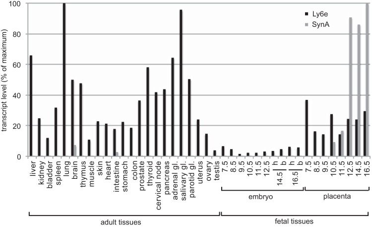
Syncytin genes are envelope genes of retroviral origin that have been exapted for a role in placentation. They are involved in the formation of a syncytial structure (the syncytiotrophoblast) at the fetomaternal interface via their fusogenic activity. The mouse placenta is unique among placental mammals since the fetomaternal interface comprises two syncytiotrophoblast layers (ST-I and ST-II) instead of one, as observed in humans and all other hemochorial placentae. Each layer specifically expresses a distinct mouse syncytin, namely, syncytin-A (SynA) for ST-I and syncytin-B (SynB) for ST-II, which have been shown to be essential to placentogenesis and embryo survival. Their cognate cellular receptors, which are necessary to mediate cell-cell fusion and syncytiotrophoblast formation, are still unknown. By devising a sensitive method that combines a cell-cell fusion assay with the screening of a mouse cDNA library, we succeeded in identifying the glycosylphosphatidylinositol (GPI)-anchored membrane protein lymphocyte antigen 6E (Ly6e) as a candidate receptor for SynA. Transfection of cells with the cloned receptor led to their fusion to cells expressing SynA, with no cross-reactive fusion activity with SynB. Knocking down Ly6e greatly reduced SynA-induced cell fusion, thus suggesting that Ly6e is the sole receptor for SynA in vivo Interaction of SynA with Ly6e was further demonstrated by a competition assay using the soluble ectodomain of Ly6e. Finally, reverse transcription-quantitative PCR (RT-qPCR) analysis of Ly6e expression on a representative panel of mouse tissues shows that it is significantly expressed in the mouse placenta together with SynA.IMPORTANCE Syncytin genes are envelope genes of endogenous retroviruses, co-opted for a physiological function in placentation. Syncytins are fusogenic proteins that mediate cell-cell fusion by interacting with receptors present on the partner cells. Here, by devising a sensitive in vitro fusion assay that enables the high-throughput screening of normalized cDNA libraries, we identified the long-sought receptor for syncytin-A (SynA), a mouse syncytin responsible for syncytiotrophoblast formation at the maternofetal interface of the mouse placenta. This protein, Ly6e (lymphocyte antigen 6E), is a GPI-anchored membrane protein, and small interfering RNA (siRNA) experiments targeting its deletion as well as a decoy assay using a recombinant soluble receptor show that Ly6e is the necessary and sufficient partner of SynA. Its profile of expression is consistent with a role in both ancestral endogenization of a SynA founder retrovirus and present-day placenta formation. This study provides a powerful general method to identify genes involved in cell-cell fusion processes.

Keywords: endogenous retrovirus; envelope protein; mouse; placenta; receptor; syncytin.


Figures

FIG 1
FIG 1
Syncytin-mediated cell-cell fusion assay: detection, quantification, and sensitivity. (A) α-Complementation of β-galactosidase was tested in A23 cells by independently transfecting or cotransfecting Galα and Galω expression vectors and analyzing β-galactosidase activity after fixation and X-Gal staining at 24 h posttransfection. (B) Syn1 and SynA fusogenicity was tested in A23 cells by a cell-cell fusion assay as shown by the scheme. (C) Stable expression of Galα in an A23-Galα cell clone was assessed by X-Gal staining at 24 h after transfection of Galω expression vector. NT, not transfected. (D and E) ASCT2/Syn1 partnership was tested with various ratios of ASCT2 expression vector mixed with a control empty vector by a cell-cell fusion assay as described for panel B using A23 and A23-Galα cells. Representative images of at least three independent experiments are shown in panels A to D. (E) The number of syncytia and the mean area of a syncytium were assessed using ImageJ software on microscope images. Data are the means ± SEM (four independent experiments; *, P < 0.05; **, P < 0.01, Student's t test). a.u., arbitrary units.
FIG 2
FIG 2
cDNA expression library screening to identify the syncytin-A partner of fusion. (A) Scheme of the screening of a mouse cDNA expression library by cell-cell fusion, which resulted in the identification of Ly6e as the potential receptor of SynA. Representative cell images of each step of the screening are shown with the range of syncytium numbers per well. (B) Scheme of mouse immature and mature Ly6e protein. In the mature form, the extracellular domain (in red) is attached to the plasma membrane by a GPI anchor on the amino acid residue boxed in the immature form after cleavage of the signal peptide (in gray) and the propeptide sequences (in white).
FIG 3
FIG 3
Ly6e is necessary and sufficient for syncytin-A mediated cell-cell fusion and is not a syncytin-B receptor. The cell-cell fusion assay was used to assess the role of Ly6e in cellulo and the specificity of the SynA/Ly6e partnership. (A) The specificity of the Ly6e partnership with SynA and not SynB was assessed by transfecting the proteins as indicated in A23 and A23-Galα cells and analyzing the formation of syncytia (presence or absence) after coculture of the two groups of cells and subsequent X-Gal staining. (B to E) The role of endogenous Ly6e in SynA-induced cell fusion was tested in human 293T embryonic kidney cells and in mouse C2C12 undifferentiated myoblasts, as indicated, by knocking down Ly6e by specific siRNAs (siLy6e) and analyzing the number of SynA-induced syncytia in comparison to those formed with nontargeting (siNT) or ASCT2 (siASCT2)-specific siRNAs. siRNA efficiency was verified by RT-qPCR (B and D), and the number of syncytia per well was analyzed by ImageJ (C) or by visual counting (E). Representative images of at least three independent experiments are shown. Data are the means ± SEM (at least three independent experiments; *, P < 0.05; **, P < 0.01, Student's t test).
FIG 4
FIG 4
Inhibition of syncytin-A mediated cell-cell fusion by soluble Ly6e recombinant proteins. (A) Scheme of mouse full-length immature Ly6e and soluble Fc- and His-tagged Ly6e. The signal peptide is in gray, the propeptide is in white, the extracellular domain is in red, the tags are in green, and the amino acid to which the GPI anchor is attached is boxed. (B) Supernatants of 293T cells untransfected or transfected with the indicated expression vectors were analyzed by Western blotting using anti-IgG and anti-His antibodies. NT, not transfected. (C) Scheme of the expected results of coculturing a first group of 293T cells overexpressing SynA and Galω and a second group overexpressing either Galα alone or Galα and a secreted soluble form of Ly6e. In the absence of soluble Ly6e, endogenous Ly6e interacts with SynA, which leads to cell-cell fusion and blue-stained syncytia; conversely, the presence of secreted soluble Ly6e will compete with endogenous Ly6e for SynA interaction, resulting in decreased formation of blue syncytia. (D) The decoy effect of soluble Ly6e (Ly6e-His and Ly6e-Fc) in comparison to an irrelevant Fc-tagged soluble receptor (EFNA4-Fc), the full-length Ly6e, or a control empty vector (ctrl) was investigated by a cell-cell fusion assay using 293T cells transfected with the indicated expression vectors. Representative images of X-Gal-stained cells are shown.
FIG 5
FIG 5
Expression profile of Ly6e and syncytin-A in a panel of adult and fetal mouse tissues: coexpression in the placenta. Ly6e and SynA transcript levels were assessed by real-time quantitative RT-PCR on tissues of adult male mice (liver to parotid gland [gl] and testis), adult female mice (uterus and ovary), embryo head and body, and placenta at indicated days of embryonic development. The embryo head (h) and body (b) were analyzed separately whenever possible. Tissue samples were analyzed in duplicate, and transcript levels were normalized relative to the amount of the RPLP0 housekeeping gene transcript. Transcript levels are represented as the percentage of maximal levels, reached in adult lung for Ly6e and in the placenta at embryonic day 16.5 for SynA.

References

    1. Lander ES, Linton LM, Birren B, Nusbaum C, Zody MC, Baldwin J, Devon K, Dewar K, Doyle M, FitzHugh W, Funke R, Gage D, Harris K, Heaford A, Howland J, Kann L, Lehoczky J, LeVine R, McEwan P, McKernan K, Meldrim J, Mesirov JP, Miranda C, Morris W, Naylor J, Raymond C, Rosetti M, Santos R, Sheridan A, Sougnez C, Stange-Thomann Y, Stojanovic N, Subramanian A, Wyman D, Rogers J, Sulston J, Ainscough R, Beck S, Bentley D, Burton J, Clee C, Carter N, Coulson A, Deadman R, Deloukas P, Dunham A, Dunham I, Durbin R, French L, Grafham D, et al. . 2001. Initial sequencing and analysis of the human genome. Nature 409:860–921. doi:10.1038/35057062. - DOI - PubMed
    1. Waterston RH, Lindblad-Toh K, Birney E, Rogers J, Abril JF, Agarwal P, Agarwala R, Ainscough R, Alexandersson M, An P, Antonarakis SE, Attwood J, Baertsch R, Bailey J, Barlow K, Beck S, Berry E, Birren B, Bloom T, Bork P, Botcherby M, Bray N, Brent MR, Brown DG, Brown SD, Bult C, Burton J, Butler J, Campbell RD, Carninci P, Cawley S, Chiaromonte F, Chinwalla AT, Church DM, Clamp M, Clee C, Collins FS, Cook LL, Copley RR, Coulson A, Couronne O, Cuff J, Curwen V, Cutts T, Daly M, David R, Davies J, Delehaunty KD, Deri J, Dermitzakis ET, et al. . 2002. Initial sequencing and comparative analysis of the mouse genome. Nature 420:520–562. doi:10.1038/nature01262. - DOI - PubMed
    1. Lavialle C, Cornelis G, Dupressoir A, Esnault C, Heidmann O, Vernochet C, Heidmann T. 2013. Paleovirology of “syncytins,” retroviral env genes exapted for a role in placentation. Philos Trans R Soc B Biol Sci 368:20120507. doi:10.1098/rstb.2012.0507. - DOI - PMC - PubMed
    1. Blaise S, de Parseval N, Benit L, Heidmann T. 2003. Genomewide screening for fusogenic human endogenous retrovirus envelopes identifies syncytin 2, a gene conserved on primate evolution. Proc Natl Acad Sci U S A 100:13013–13018. doi:10.1073/pnas.2132646100. - DOI - PMC - PubMed
    1. Blond JL, Lavillette D, Cheynet V, Bouton O, Oriol G, Chapel-Fernandes S, Mandrand B, Mallet F, Cosset FL. 2000. An envelope glycoprotein of the human endogenous retrovirus HERV-W is expressed in the human placenta and fuses cells expressing the type D mammalian retrovirus receptor. J Virol 74:3321–3329. doi:10.1128/JVI.74.7.3321-3329.2000. - DOI - PMC - PubMed

Publication types