. 2017 Aug 24;91(18):e00574-17.
doi: 10.1128/JVI.00574-17. Print 2017 Sep 15.

Mutational Analysis of Lassa Virus Glycoprotein Highlights Regions Required for Alpha-Dystroglycan Utilization

Affiliations

Mutational Analysis of Lassa Virus Glycoprotein Highlights Regions Required for Alpha-Dystroglycan Utilization

Marissa Acciani et al. J Virol. .

Abstract

Lassa virus (LASV) is an enveloped RNA virus endemic to West Africa and responsible for severe cases of hemorrhagic fever. Virus entry is mediated by the glycoprotein complex consisting of a stable-signal peptide, a receptor-binding subunit, GP1, and a viral-host membrane fusion subunit, GP2. Several cellular receptors can interact with the GP1 subunit and mediate viral entry, including alpha-dystroglycan (αDG) and lysosome-associated membrane protein 1 (LAMP1). In order to define the regions within GP1 that interact with the cellular receptors, we implemented insertional mutagenesis, carbohydrate shielding, and alanine scanning mutagenesis. Eighty GP constructs were engineered and evaluated for GP1-GP2 processing, surface expression, and the ability to mediate cell-to-cell fusion after low-pH exposure. To examine virus-to-cell entry, 49 constructs were incorporated onto vesicular stomatitis virus (VSV) pseudoparticles and transduction efficiencies were monitored in HAP1 and HAP1-ΔDAG1 cells that differentially produce the αDG cell surface receptor. Seven constructs retained efficient transduction in HAP1-ΔDAG1 cells yet poorly transduced HAP1 cells, suggesting that they are involved in αDG utilization. Residues H141, N146, F147, and Y150 cluster at the predicted central core of the trimeric interface and are important for GP-αDG interaction. Additionally, H92A-H93A, 150HA, 172HA, and 230HA displayed reduced transduction in both HAP1 and HAP1-ΔDAG1 cells, despite efficient cell-to-cell fusion activity. These mutations may interfere with interactions with the endosomal receptor LAMP1 or interfere at another stage in entry that is common to both cell lines. Insight gained from these data can aid in the development of more-effective entry inhibitors by blocking receptor interactions.IMPORTANCE Countries in which Lassa virus is endemic, such as Nigeria, Sierra Leone, Guinea, and Liberia, usually experience a seasonal outbreak of the virus from December to March. Currently, there is neither a preventative vaccine nor a therapeutic available to effectively treat severe Lassa fever. One way to thwart virus infection is to inhibit interaction with cellular receptors. It is known that the GP1 subunit of the Lassa glycoprotein complex plays a critical role in receptor recognition. Our results highlight a region within the Lassa virus GP1 protein that interacts with the cellular receptor alpha-dystroglycan. This information may be used for future development of new Lassa virus antivirals.

Keywords: arenavirus; receptor binding; virus entry.


Figures

FIG 1
FIG 1
Subunits and structure of the Lassa virus glycoprotein complex. (A) The LASV glycoprotein complex consists of the membrane-integrated stable-signal peptide (SSP) (blue), the GP1 subunit (purple), and the noncovalently attached GP2 subunit (green). The GPC is cleaved by the signal peptidase (red arrow) and SKI-1/S1P (yellow arrow) during protein processing. (B) Cartoon of the SSP, GP1, and GP2 heterotrimer complex in the lipid bilayer. (C) Trimeric LASV GP1-GP2 crystal structure, viewed from the top down; GP1 is in purple and GP2 in green (PDB 5vk2) (32). (D) LASV GP1-GP2 crystal structure, side view (PDB 5vk2). The engineered GP1 constructs are color coded as follows: pink, glycosylation site removals and additions; orange, HA-tagged sites; yellow, alanine scanning of charged residues; blue, alanine scanning of hydrophobic residues; red, additional targeted residues. Note that many of the targeted residues were found in regions of the structure that did not crystallize. All structures were rendered with PyMol.
FIG 2
FIG 2
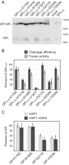
Processing and functional characteristics of surface-expressed GP1 N-glycosylation sites. (A) Vero cells were transfected with the indicated FLAG-tagged LASV GP variant or the negative control. After 36 h, cells were subjected to surface biotinylation. Surface-expressed biotinylated proteins were concentrated using streptavidin Sepharose beads. Precipitated proteins were separated by SDS-PAGE. Immunoblot assays were carried out to detect LASV GP surface-expressed protein using an anti-FLAG antibody, M2. The immunoblot shown is representative of four trials. (B) Microphotographs of Vero cells cotransfected with plasmid DNA encoding LASV GP construct and GFP. Cell-to-cell fusion was assessed 3 h following low-pH-medium shock; magnification, ×20. Representative fields of view are shown. (C) Fusion data for each construct was quantified by counting unfused cells and comparing the numbers to those in mock-transfected wells. Quantified fusion data for each construct were normalized to LASV wt-GPC-3xFLAG. Cleavage efficiency was normalized to FLAG-tagged GP using densitometry analysis. (D) Parental GP transduction efficiency in HAP1 and HAP1-ΔDAG1 cells. VSVΔG-GFP pseudoparticles containing LASV GP were added to both cells, and the GFP-positive cells were enumerated in a flow cytometer. The percentage of the cell population that was GFP positive is shown. (E) VSVΔG-GFP pseudoparticles containing LASV GP or N-glycosylation mutants were used to transduce HAP1 and HAP1-ΔDAG1 cells. The GFP-positive cells were enumerated in a flow cytometer. Transduction efficiencies were normalized to parental LASV GP particle transduction values in each respective cell type. All data are based on the averages and standard errors of the means from at least three replicate experiments.
FIG 3
FIG 3
Functional analysis of GP1 containing engineered N-linked glycosylation sites. (A) Surface-expressed GP of N-glycan mutants and immunoblot analysis using anti-FLAG antibody M2 for detection. (B) Cleavage efficiency and cell-to-cell fusion data. (C) Transduction of HAP1 and HAP1-ΔDAG1 cells using VSV-pseudotyped particles. All data are based on the averages and standard errors of the means from at least three replicate experiments.
FIG 4
FIG 4
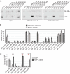
Insertional mutagenesis of LASV GPC blocks entry in specific cell lines. (A) Surface-expressed HA-tagged mutants and immunoblot analysis using anti-FLAG antibody M2. (B) Cleavage efficiency and cell-to-cell fusion data. (C) Transduction of HAP1 and HAP1-ΔDAG1 cells using VSV-pseudotyped particles. All data are based on the averages and standard errors of the means from at least three replicate experiments. ***, P < 0.001.
FIG 5
FIG 5
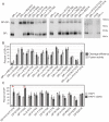
Mutating hydrophobic GP1 residues impedes protein processing. (A) Surface-expressed hydrophobic mutants and immunoblot analysis using anti-FLAG antibody M2. (B) Cleavage efficiency and cell-to-cell fusion data. (C) Transduction of HAP1 and HAP1-ΔDAG1 cells using VSV-pseudotyped particles. All data are based on the averages and standard errors of the means from at least three replicate experiments. The horizontal blue bar highlights the construct that transduced HAP1-ΔDAG1 in a fashion similar to that of parental GP yet showed a defect in HAP1 entry.
FIG 6
FIG 6
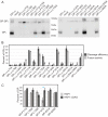
Charged GP1 residues are required for efficient HAP1 entry. (A) Surface-expressed charged mutants and immunoblot analysis using anti-FLAG antibody M2. (B) Cleavage efficiency and fusion ability data. (C) Transduction of HAP1 and HAP1-ΔDAG1 cells using VSV-pseudotyped particles. All data are based on the averages and standard errors of the means from at least three replicate experiments. The horizontal blue bars highlight the constructs that transduced HAP1-ΔDAG1 more than twice as efficiently as HAP1 cells.
FIG 7
FIG 7
Residues involved with the GP1 trimer core are critical for αDG interaction. (A) Surface-expressed targeted GP1 mutants and immunoblot analysis using anti-FLAG antibody M2. (B) Cleavage efficiency and fusion ability data. (C) Transduction of HAP1 and HAP1-ΔDAG1 cells using VSV-pseudotyped particles. All data are based on the averages and standard errors of the means from at least three replicate experiments. *, P < 0.05; **, P < 0.01.
FIG 8
FIG 8
GP constructs are efficiently incorporated onto VSV particles. Vero cells were used to produce VSV pseudoparticles. Cell supernatants, containing pseudotyped particles, were collected and precipitated with TCA to determine the level of LASV GP incorporation into VSV envelopes. GP constructs demonstrating low HAP1 transduction or GP constructs that were unable to transduce both cell types were tested. Precipitated proteins were separated by SDS-PAGE and immunoblotted for LASV GP2 (monoclonal antibody 12.4D) and VSV-M (monoclonal antibody 23H12).
FIG 9
FIG 9
Identification and mapping residues implicated in LASV receptor binding and viral entry. (A) To directly examine GP1 mutant binding, or lack of binding, to αDG, we performed a coimmunoprecipitation assay. αDG-coated beads were incubated with VSV-pseudotyped particles containing either parental or mutant LASV GP. All proteins interacting with the αDG-coated beads were concentrated and separated by SDS-PAGE. While parental GP and GP-H230Y were able to bind to αDG, GP-H141A-F147A and GP-R248A-R250A were not. (B) LASV GP constructs exhibiting reduced entry into HAP1 cells were mapped onto the LASV prefusion crystal structure (PDB 5vk2; GP1 is shown in purple, and GP2 is shown in green). Regions that are significantly involved in αDG interactions are shown in red (H141, N146, F147, and Y150). Regions that also demonstrated reduced HAP1 entry are shown in orange (R248 and R250) and yellow (K125, K126, and W227). The histidine triad is shown in pink (H92, H93, and H230). Residues are color coded to match Table 1. Both a cartoon structure and surface rendering of the structure demonstrate that the αDG binding site is located in a cavity on the top of the trimer. (C) Sequence alignment of Old World arenaviruses that use αDG. Regions in LCMV involved in αDG binding are shown in blue, while the regions that we have implicated for LASV entry are shown in red. R248 and R250 are highlighted in orange. The histidine triad implicated in LAMP1 interaction is in pink.

References

    1. Frame JD, Baldwin JM Jr, Gocke DJ, Troup JM. 1970. Lassa fever, a new virus disease of man from West Africa. I. Clinical description and pathological findings. Am J Trop Med Hyg 19:670–676. - PubMed
    1. Gunther S, Lenz O. 2004. Lassa virus. Crit Rev Clin Lab Sci 41:339–390. doi:10.1080/10408360490497456. - DOI - PubMed
    1. Mofolorunsho KC. 2016. Outbreak of lassa fever in Nigeria: measures for prevention and control. Pan Afr Med J 23:210. doi:10.11604/pamj.2016.23.210.8923. - DOI - PMC - PubMed
    1. Fichet-Calvet E, Rogers DJ. 2009. Risk maps of Lassa fever in West Africa. PLoS Negl Trop Dis 3:e388. doi:10.1371/journal.pntd.0000388. - DOI - PMC - PubMed
    1. Ogbu O, Ajuluchukwu E, Uneke CJ. 2007. Lassa fever in West African sub-region: an overview. J Vector Borne Dis 44:1–11. - PubMed

Publication types

MeSH terms