. 2017 Sep 1;118(3):1871-1887.
doi: 10.1152/jn.01166.2015. Epub 2017 Jul 5.

Envelope contributions to the representation of interaural time difference in the forebrain of barn owls

Affiliations

Envelope contributions to the representation of interaural time difference in the forebrain of barn owls

Philipp Tellers et al. J Neurophysiol. .

Abstract

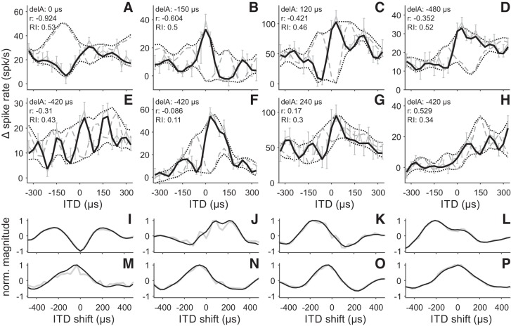
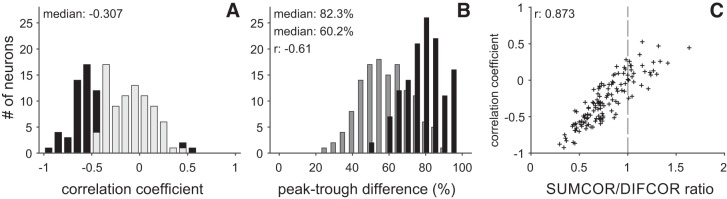
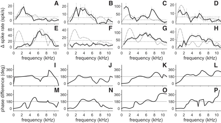
Birds and mammals use the interaural time difference (ITD) for azimuthal sound localization. While barn owls can use the ITD of the stimulus carrier frequency over nearly their entire hearing range, mammals have to utilize the ITD of the stimulus envelope to extend the upper frequency limit of ITD-based sound localization. ITD is computed and processed in a dedicated neural circuit that consists of two pathways. In the barn owl, ITD representation is more complex in the forebrain than in the midbrain pathway because of the combination of two inputs that represent different ITDs. We speculated that one of the two inputs includes an envelope contribution. To estimate the envelope contribution, we recorded ITD response functions for correlated and anticorrelated noise stimuli in the barn owl's auditory arcopallium. Our findings indicate that barn owls, like mammals, represent both carrier and envelope ITDs of overlapping frequency ranges, supporting the hypothesis that carrier and envelope ITD-based localization are complementary beyond a mere extension of the upper frequency limit.NEW & NOTEWORTHY The results presented in this study show for the first time that the barn owl is able to extract and represent the interaural time difference (ITD) information conveyed by the envelope of a broadband acoustic signal. Like mammals, the barn owl extracts the ITD of the envelope and the carrier of a signal from the same frequency range. These results are of general interest, since they reinforce a trend found in neural signal processing across different species.

Keywords: auditory arcopallium; carrier; envelope; extracellular recordings; sound localization.


Figures

Fig. 1.
Fig. 1.
Noise stimuli. A: broadband white noise as used in the experiments (0.5–25 kHz). B: band-pass filtered noise. Because of filtering at the level of the cochlea, broadband noise stimuli are split into 1/3-octave-wide frequency bands. The filtering produces a periodic carrier and an aperiodic envelope. C: close up of the carrier (car0) and the envelope (env0) of the band-pass filtered noise shown in B. Besides the band-pass noise, the carrier (car180) and envelope (env180) of a 180° phase shifted noise are also shown. The stimulus carrier is phase sensitive. A 180° phase shift causes an inversion of the carrier (compare gray lines). By contrast, the stimulus envelope is unaffected by the phase shift (compare black lines). The envelope is phase invariant.
Fig. 2.
Fig. 2.
Envelope extraction and delay asymmetry. A: pair of modeled NDFs (NDF0, NDF180). The NDF envelope functions were extracted by smoothing the min and max functions. The min function is given by the lowest response of the NDF pair at each ITD, while the max function represents the highest response at each ITD. The NDF envelope functions were aligned with the local extrema of the 2 NDFs. Shifting the envelope functions did not affect the envelope’s shape or the output of the cross-correlation analysis. B: cross-correlation function of the 2 NDF envelope functions (black line). The upper and lower envelopes were cross-correlated to determine the delay asymmetry. The delay asymmetry is defined as the ITD shift that maximized the anticorrelation between the upper and lower envelope functions. The minimum of the cross-correlation functions gives the ITD shift that maximizes the anticorrelation and thus the delay asymmetry. The gray line shows the cross-correlation function of the min and max functions. The cross-correlation function exhibited an oscillating high-pass element that is a sampling artifact and is due to the fact that we recorded only 2 NDFs (0 and 180°). Note that the 2 cross-correlation functions are similar with the exception of the oscillating high-pass component. Both functions exhibit the minimum at 0 µs.
Fig. 3.
Fig. 3.
Carrier and envelope elements in NDFs. A: modeled response of a solely carrier-sensitive neuron. The NDF0 function (solid black line) was generated by cross-correlating 2 copies of the same carrier signal (car0/car0), similar to the one shown in Fig. 1, B and C. The NDF180 (dashed gray line), was created by cross-correlating a carrier (Fig. 1C; car0) and its 180° phase-shifted counterpart (Fig. 1C; car180). The black dotted lines represent the envelope functions of the 2 NDFs. The delay asymmetry (delA) and the correlation (r) of the NDF pair are given at top left of each panel. A neuron that is solely driven by the stimulus carrier has a correlation coefficient of −1 and no delay asymmetry. B: modeled response of a solely envelope-sensitive neuron. The functions and values are the same as in A with the exception that the NDF0 and NDF180 were created by cross-correlating the stimulus envelopes (Fig. 1C, black lines; env0 and env180) instead of the carriers. The stimulus envelope is phase invariant. Thus the NDF0 and NDF180 are similar for a solely envelope-driven neuron. The correlation of the 2 NDFs is close to 1. Note that the NDF180 and the 2 NDF envelopes were shifted to allow an unobstructed view on all 4 curves. C: modeled response of a partly envelope-sensitive neuron. The 2 NDFs were created by mixing the NDF functions shown in A and B in equal ratio. The correlation of a carrier and envelope driven neuron is intermediate, while the delay asymmetry is nonzero. D: rectified responses of a solely carrier-sensitive model neuron. The NDFs are rectified versions of the NDFs shown in A. Rectification decreases anticorrelation (r: −0.53 compared with −1 in A) but does not introduce a delay asymmetry (delA: 0 µs). E–H: cross-correlation functions of the 4 modeled neurons (black lines). The cross-correlation of the upper and lower envelopes was used to determine the neuron’s delay asymmetry. The delay asymmetry is defined as the ITD shift that maximizes the anticorrelation between the upper and lower envelope functions, i.e., the minimum of the cross-correlation function. The panels also show the cross-correlation functions of the min and max functions (gray line) (compare Fig. 2B). I–L: DIFCOR functions of the modeled responses shown in A–D. DIFCOR functions are created by subtracting the NDF180 from the NDF0. The DIFCOR represents the phase-sensitive component of the response. M–P: SUMCOR functions of the modeled NDFs. SUMCOR functions are the sum of the 2 NDFs and represent the phase-tolerant response component.
Fig. 4.
Fig. 4.
Effect of the envelope and its ITD on delay asymmetry. A–I: modeled NDFs with varying carrier and envelope contributions (black line: NDF0; dashed gray line: NDF180). Envelope contributions increases from top (10%) to bottom (30%), while the best ITD of the envelope component increases from left to right (30 µs, 180 µs and, 330 µs). The best ITD is defined as the ITD that elicits the highest response. The best ITD of the carrier component was kept at 30 µs for all modeled NDFs. The delay asymmetry (DA) and the correlation (r) are given at top left of each panel. Upper and lower NDF envelopes are shown as black dotted lines. J–L: cross-correlation functions computed for increasing envelope contributions (10–30% in 5% steps) and varying best ITDs of the envelope component (J: 30 µs, K: 180 µs; L: 330 µs). Darker curves correspond to smaller envelope contributions, and lighter curves correspond to bigger envelope contribution. Solid lines represent the cross-correlation function for the neurons shown in A–I with 10%, 20%, and 30% envelope contributions, while the 2 dashed lines show the cross-correlation functions for neurons with 15% and 25% envelope contributions (NDFs not shown). Note that for all 3 ITD configurations the position of the cross-correlation’s minimum shifts from 0 µs to more extreme values with increasing envelope contribution.
Fig. 5.
Fig. 5.
Typical NDFs of AAr neurons. A–H: NDFs recorded with correlated (NDF0; black solid lines) and anticorrelated, i.e., 180° phase-shifted (NDF180; gray dashed lines) noise of 8 AAr neurons. NDFs were low-pass filtered at 10 kHz to remove high-frequency noise. For the NDF0, we also included the raw, unfiltered data (light gray lines). Error bars give SE of the change (Δ) in spike rate. Upper and lower NDF envelope functions are shown as black dotted lines. The correlation (r) between the 2 NDFs, the delay asymmetry (delA), and the rectification index (RI) are given at top left of each panel. Note that the correlation of the neurons increases from A to H. I–P: cross-correlation functions of the NDF envelopes shown in A–H (black lines). The position of the cross-correlation’s minimum gives the neuron’s delay asymmetry. Gray lines depict the cross-correlation of the unsmoothed min and max functions (not shown, see Fig. 2).
Fig. 6.
Fig. 6.
Identification of envelope-sensitive neurons. A: distribution of delay asymmetries. Gray dashed line marks the 30 µs cutoff used for classifying neurons. Neurons with a magnitude of delay asymmetry that exceeded 30 µs were classified as envelope sensitive; 137 of the 146 neurons exhibited a magnitude of delay asymmetry of at least 60 µs. B: distribution of rectification indexes. Most neurons exhibited intermediate rectification indexes between 0.2 and 0.6, with a population median of 0.423. Gray dashed line in the distribution of the rectification indexes marks the 0.2 threshold. The 5 neurons that exhibited a rectification index below 0.2 and the 9 neurons with a magnitude of delay asymmetry below 60 µs were removed from further analysis. The remaining 132 neurons of the data set carried envelope-sensitive elements in their responses.
Fig. 7.
Fig. 7.
DIFCOR and SUMCOR functions. A–H: DIFCOR functions of the same AAr neurons shown in Fig. 5. The DIFCOR was created by subtracting the NDF180 from the NDF0. It represents the phase-sensitive component of the NDFs. I–P: SUMCOR functions of the neurons shown in Fig. 5 (black dashed lines). SUMCOR functions are the sum of the 2 NDFs and represent the phase-invariant response component. DIFCOR and SUMCOR functions were normalized by dividing them by their maximum possible peak-trough difference. The maximum possible peak-trough difference of a neurons is set by the sum of the peak-trough differences of its NDF0 and NDF180. The peak-trough difference (PT) and the best ITD of the DIFCOR and SUMCOR functions are given at top left of each panel. The best ITD of a function is given by the ITD that elicits the highest response. SUMCOR functions were low-pass filtered to remove high-pass components in the response (gray lines) before determination of best ITD. The high-pass artifact arises because SUMCOR and DIFCOR functions are computed from a set of NDFs with only 2 different phase configurations, 0 and 180° phase-shifted.
Fig. 8.
Fig. 8.
Phase sensitivity and envelope contributions. A: distribution of correlation coefficients. Significant coefficients are shown in black (Pearson’s r, P < 0.05). Nonsignificant correlation coefficients are shown in light gray. B: distribution of peak-trough differences of the DIFCOR (black) and SUMCOR (gray) functions. The DIFCOR functions exhibit a significantly higher peak-trough difference than the SUMCOR functions (Wilcoxon signed-rank test, P < 0.001). The peak-trough differences of the 2 functions were negatively correlated (r: −0.61; Pearson’s r, P < 0.001). C: correlation between the correlation coefficient and the SUMCOR-to-DIFCOR ratio (SUMCOR/DIFCOR). SUMCOR/DIFCOR was calculated by dividing the peak-trough difference of the SUMCOR by the peak-trough difference of the DIFCOR function. A SUMCOR/DIFCOR above 1 indicates an envelope-dominated response, while a ratio below 1 corresponds to a carrier-dominated response (dashed gray line). Correlation coefficient and SUMCOR/DIFCOR were significantly correlated (r: 0.873, Pearson’s r, P < 0.001).
Fig. 9.
Fig. 9.
Best ITD of DIFCOR and SUMCOR. A: distribution of the best ITDs of the DIFCOR (black bars) and SUMCOR (gray bars) functions. The best ITD is defined as the ITD that elicits the highest response. Both DIFCOR and SUMCOR exhibit mostly positive best ITDs with a peak close to 0. The SUMCOR functions have significantly bigger best ITDs than the DIFCOR functions (Wilcoxon signed-rank test, P < 0.001). The best ITDs of the 2 functions are only weakly correlated (r = 0.27, Pearson’s r, P < 0.001). B: distribution of ITD differences. The best ITD difference is given by the absolute difference between the best ITD of the DIFCOR and the best ITD of the SUMCOR. The majority of AAr neurons (88 of 132) exhibited a best ITD difference of at least 60 µs.
Fig. 10.
Fig. 10.
Frequency-response functions and phase difference spectra. A–H: frequency-response functions of the 8 example neurons shown in Fig. 5 (black solid lines). Error bars give SE of the change in spike rate. Gray dashed lines represent the power spectrum of the neurons’ NDF0. Power and phase spectra were computed with a discrete Fourier transformation. The 1/3 magnitude level of the frequency spectra is marked by the black dotted line. Only those parts of the power spectrum that exceeded the 1/3 magnitude threshold were considered to carry energy. I–P: phase difference spectra of the same 8 example neurons (black lines). Phase difference spectra were created by subtracting the phase spectrum of the NDF180 from the phase spectrum of the NDF0. A phase difference of ±180 indicates phase sensitivity (dotted lines), while a phase difference of 0 or 360 indicates phase invariance.
Fig. 11.
Fig. 11.
Mean phase difference spectrum. A: the mean phase difference spectrum (black line) was created by averaging the phase difference spectra of the 132 AAr neurons that exhibited envelope sensitivity. Only the section of the phase spectrum where the neuron’s power spectrum reached at least 1/3 of its maximum magnitude was used for averaging (see dotted lines in Fig. 10). Gray line shows the vector strength of the averaged phase spectrum. B–D: polar plots show the neurons’ phase differences (gray plus signs) at 3 different frequencies (black x-signs in A). The direction of the black line in the center of each polar plot gives the mean phase spectrum at these frequencies, while its length corresponds to the vector strength. The vector strength is a measure for the similarity of the phase difference spectrum across the population. A vector strength of 1 indicates that all neurons have the same phase difference, while a vector strength of 0 indicates a lack of correlation across the population. Values for the averaged phase difference (avg) and the vector strength (VS) are shown at top of each panel.
Fig. 12.
Fig. 12.
AAr responses to high-pass filtered noise. A–D: NDF0 (black lines) and NDF180 (gray dashed lines) of the 4 neurons shown in Fig. 5, A, D, F, and H. NDFs were recorded using high-pass filtered noise stimuli. Upper and lower envelope functions are shown as black dotted lines. Delay asymmetry (delA) and correlation coefficients (r) are given at top left of each panel. The cutoff frequency for the high-pass noise stimuli was chosen individually for each neuron depending on its frequency response function (compare Fig. 10). Cutoff frequencies ranged from 2.5 to 5.5 kHz. E–H: cross-correlation functions of the NDF envelope functions shown in A–D (black lines). The position of the cross-correlation’s minimum determines the neurons delay asymmetry. Gray lines depict the cross-correlation functions of the min and max functions (not shown; see Fig. 2). I–L: power spectra of the NDF0 shown in A–D. Power spectra were computed with fast Fourier transformation algorithm.
Fig. 13.
Fig. 13.
Envelope contributions in responses to high-pass filtered stimuli. A: distribution of delay asymmetries for 18 of the 21 NDF pairs recorded with high-pass filtered noise stimuli. The missing 3 neurons were excluded before the analysis because their maximum response rate did not exceed 10 spk/s (<1 spike per stimulation) (Fig. 12A). Seventeen of the remaining 18 neurons had a magnitude of delay asymmetry of at least 60 µs. Gray dashed line shows the 30 µs threshold used for identifying envelope sensitivity. B: distribution of the correlation coefficients of the 17 envelope-sensitive neurons. C: distribution of peak-trough differences of the 17 DIFCOR (black) and SUMCOR (gray) functions. The DIFCOR functions exhibited significantly higher peak-trough differences than the SUMCOR functions (Wilcoxon signed-rank test, P < 0.01). D: mean phase difference spectra (black line) of the 17 neurons with a delay asymmetry of at least 60 µs and their corresponding vector strength (gray line). Phase differences of ±180° indicate phase sensitivity (black dotted line), while phase differences of 0 or ±360° indicate phase invariance. E: distribution of the differences between the best ITDs of the DIFCOR and the best ITDs of the SUMCOR. The SUMCOR and DIFCOR functions exhibited a different best ITD in 16 of the 17 envelope-sensitive neurons.

References

    1. Aaronson NL, Hartmann WM. Interaural coherence for noise bands: waveforms and envelopes. J Acoust Soc Am 127: 1367–1372, 2010. doi:10.1121/1.3290991. - DOI - PMC - PubMed
    1. Agapiou JP, McAlpine D. Low-frequency envelope sensitivity produces asymmetric binaural tuning curves. J Neurophysiol 100: 2381–2396, 2008. doi:10.1152/jn.90393.2008. - DOI - PMC - PubMed
    1. Albeck Y, Konishi M. Responses of neurons in the auditory pathway of the barn owl to partially correlated binaural signals. J Neurophysiol 74: 1689–1700, 1995. - PubMed
    1. Ashida G, Carr CE. Sound localization: Jeffress and beyond. Curr Opin Neurobiol 21: 745–751, 2011. doi:10.1016/j.conb.2011.05.008. - DOI - PMC - PubMed
    1. Batra R, Kuwada S, Stanford TR. High-frequency neurons in the inferior colliculus that are sensitive to interaural delays of amplitude-modulated tones: evidence for dual binaural influences. J Neurophysiol 70: 64–80, 1993. - PubMed

Publication types

LinkOut - more resources