. 2017 Jul 5;7(1):4676.
doi: 10.1038/s41598-017-04611-x.

Linking functions: an additional role for an intrinsically disordered linker domain in the transcriptional coactivator CBP

Affiliations

Linking functions: an additional role for an intrinsically disordered linker domain in the transcriptional coactivator CBP

Sara Contreras-Martos et al. Sci Rep. .

Abstract

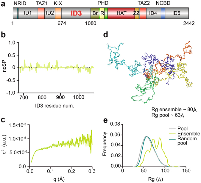
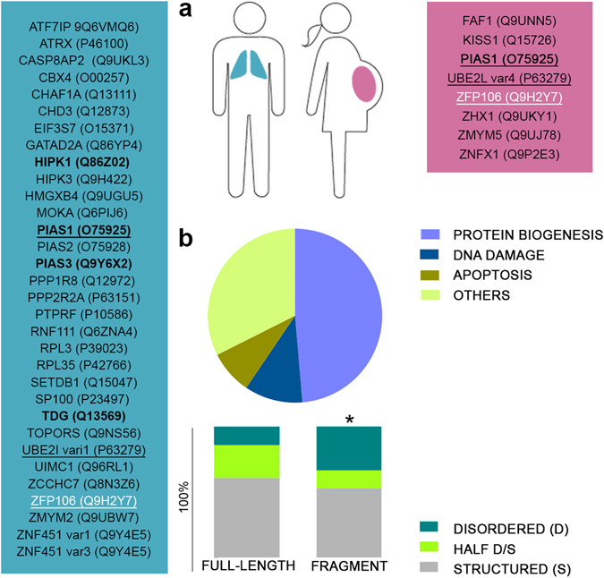
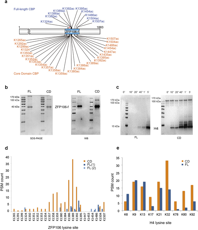
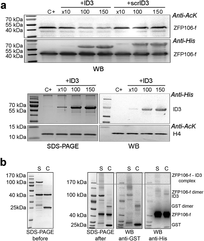
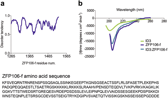
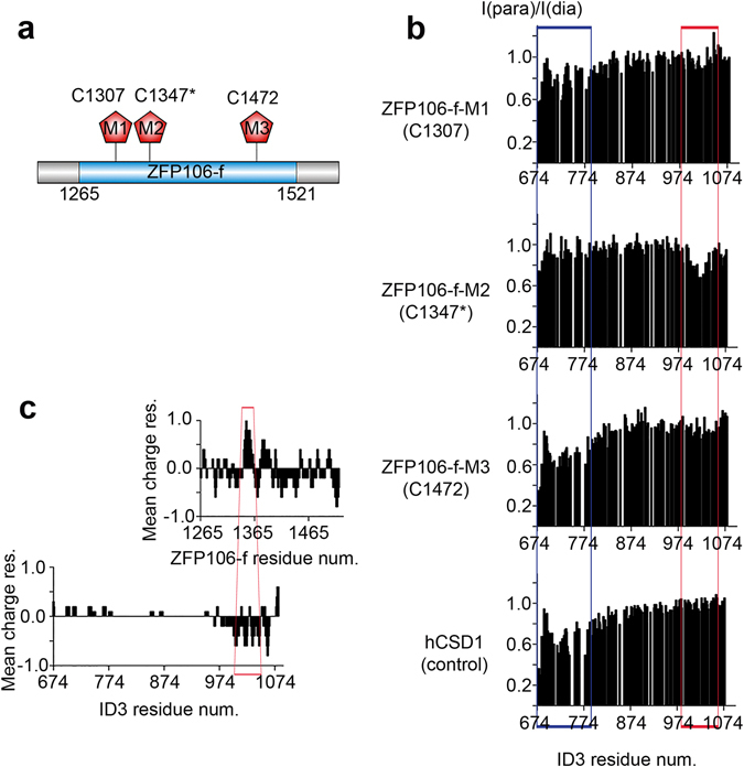
The multi-domain transcriptional coactivators CBP/p300 integrate a multitude of signaling inputs, interacting with more than 400 proteins via one or more of their globular domains. While CBP/p300 function is typically considered in terms of these structured domains, about half of the protein consists of intrinsically disordered regions (IDRs) of varying length. However, these IDRs have only been thought of as linkers that allow flexible spatial arrangement of the structured domains, but recent studies have shown that similar IDRs mediate specific and critical interactions in other proteins. To examine the roles of IDRs in CBP, we performed yeast-two-hybrid screenings of placenta and lung cancer cDNA libraries, which demonstrated that the long IDR linking the KIX domain and bromodomain of CBP (termed ID3) can potentially bind to several proteins. The RNA-binding Zinc-finger protein 106 (ZFP106) detected in both libraries was identified as a novel substrate for CBP-mediated acetylation. Nuclear magnetic resonance (NMR) spectroscopy combined with cross-linking experiments and competition-binding assays showed that the fully disordered isolated ID3 transiently interacts with an IDR of ZFP106 in a fashion that disorder of both regions is maintained. These findings demonstrate that beside the linking function, ID3 can also interact with acetylation substrates of CBP.


Conflict of interest statement

The authors declare that they have no competing interests.

Figures

Figure 1
Figure 1
Domain 3 (ID3) of CBP is fully disordered. (a) CBP domain organization. CBP globular domains (colored; Br: Bromodomain, R: Ring) are connected by intrinsically disordered regions (IDRs, grey) of different lengths. The intrinsically disordered region 3 (ID3, aa674–1080) is located between the KIX domain and bromodomain of CBP. (b) Neighbor corrected structural propensities (ncSP) obtained from experimentally measured N, C′, Cα and Cβ chemical shifts. Positive and negative values correspond to α-helical and β-sheet propensities, respectively. (c) SAXS Kratky plot also shows an overall disordered state, with a minor tendency for local secondary structure formation. (d) Ensemble of selected conformations ID3 may adopt in solution. (e) Rg distributions of a random pool of ID3 conformations (teal), of the pool of ID3 conformations biased by experimental ncSP scores (grey) and the ensemble calculated by Flexible Meccano and selected by GAJOE (green).
Figure 2
Figure 2
ID3 interacts with the transcription factor ZFP106. (a) Potential interaction partners of ID3 have been identified by a yeast 2-hybrid (Y2H) screen. Y2H interactions were classified according to the libraries in which they were detected: placenta (pink), lung cancer (blue), or both (black underlined). Four proteins have been described in previous studies to interact with one or more of the CBP’s globular domains (bold). ZFP106, selected for further studies, is marked in white. (b) Potential interactors were analyzed for their structural content (Structured: more than 60% of its sequence predicted to be structured; Disordered: more than 60% of its sequence predicted to be disordered; Half D/S: the rest; *Statistically significant preference of ID3 for binding disordered regions) and for their function. Disorder predictions were carried out with the IUPred webserver, whereas gene ontology (GO) annotations and available literature were used to classify the proteins in four main groups: Protein biogenesis (including transcription, pre-mRNA maturation and protein biosynthesis), DNA damage, Apoptosis and Others.
Figure 3
Figure 3
ZFP106 is specifically acetylated only by full-length CBP. (a) ZFP106-f was acetylated by either full-length (FL) or the catalytic core domain (CD) of CBP under comparable conditions, and the sites of acetylation were determined by MS. Specific acetylation pattern by full-length CBP (blue) is in contrast to the indiscriminate acetylation achieved by the core domain (orange). *Location of the lysine rich patch of ZFP106-f. (b) SDS-PAGE and WB developed with anti-acetylated-lysine antibody of ZFP106-f acetylated by the full-length or core domain of CBP. (c) Western blot developed with anti-acetylated-lysine antibody of histone 4 (H4) acetylated by the full-length or core domain of CBP. Time points of the reaction are presented and highlight a much higher activity for the CD compared to the FL enzyme. (d and e) The ratio of acetylated peptides corresponding to various lysines identified by MS using ZFP106-f (d) or H4 (e) as a substrate, acetylated with full-length CBP (dark blue: first MS analysis; light blue: second MS analysis) or its core domain (orange).
Figure 4
Figure 4
ID3 –ZFP106-f interaction assessed by competition binding assay and cross-linking. (a) Top: Competition assays with ZFP106-f as a substrate demonstrate that recombinant ID3 competes with ID3 in the full-length CBP for binding ZFP106-f, which results in a decrease of the ZFP106-f acetylation in the presence of an excess of ID3. However, addition of the same amount of scrambled ID3 (scrID3) has no effect. Bottom: as a control, the same experiment was run using histone 4 (H4) as a substrate; no change in H4 acetylation level could be detected after addition of an excess of ID3. Acetylation was followed by anti-Ac-Lys antibody, adding ID3 to the acetylation reaction in an excess of 10, 100 or 150 times to full-length CBP (18.5 pmol) and its effect was also compared to that of scrambled-ID3 (scrID3; control reaction) (C+: control reaction). (b) Cross-linked ZFP106-ID3 complex (S: sample reaction) is shown by the presence of high-Mw overlapping bands in the SDS-PAGE gel after cross-linking, anti-His WB and anti-GST WB (box). Under similar condition, ZFP106-GST (C: control reaction) were not cross-linked. Cross-linked complexes formed during the reaction are detailed on the right side of the WB and they have been determined by comparing signals between the anti-His and anti-GST WB (GST 25 kDa; ZFP106-f 40 kDa; GST dimer 62 kDa; ID3 100 kDa, ZFP106-f dimer 115 kDa, ZFP106-f – ID3 complex above 170 kDa).
Figure 5
Figure 5
Fuzzy interaction between the two disordered regions. (a) Top: Disorder prediction of ZFP106-f using IUPred server indicates a high disorder tendency. Bottom: The ZFP106-f amino acid sequence used for the disorder prediction. (b) Circular dichroism spectra of ID3, ZFP106-f and 1:1 ratio of ID3:ZFP106-f. The spectrum of the complex resembles the individual spectra indicating no major changes in the structural content of the two partners upon association.
Figure 6
Figure 6
CTR of ID3 specifically interacts with the middle region of ZFP106-f. (a) Representation of the ZFP106 fragment identified in the Y2H assay (blue) and the three positions (M1, M2 and M3) where a paramagnetic label was attached to the protein. (b) I (para) /I(dia) plots of the 2D 1H-15N HSQC cross-peaks intensities of ID3 in the presence of the paramagnetically tagged single-Cys ZFP106-f mutants M1, M2 and M3 showing the site of interaction within ID3 (red box). A control experiment with an unrelated IDP, domain 1 of human Calpastatin (hCSD1), labeled with the same paramagnetic probe, is also shown to outline sticky region(s) of ID3 engaged in non-specific interaction (blue box). (c) Charge distribution plots of ID3 and ZFP106-f amino acid sequences calculated with the EMBOSS website (http://www.bioinformatics.nl/cgi-bin/emboss/charge). The regions involved in the interaction are highlighted (red): the middle region of ZFP106-f close to the M2 site, and the C-terminal 100 residues (CTR) of ID3.

References

    1. Bedford DC, Brindle PK. Is histone acetylation the most important physiological function for CBP and p300? Aging (Albany NY) 2012;4:247–255. doi: 10.18632/aging.100453. - DOI - PMC - PubMed
    1. Vo N, Goodman RH. CREB-binding protein and p300 in transcriptional regulation. J Biol Chem. 2001;276:13505–13508. doi: 10.1074/jbc.R000025200. - DOI - PubMed
    1. Giles RH, Peters DJ, Breuning MH. Conjunction dysfunction: CBP/p300 in human disease. Trends Genet. 1998;14:178–183. doi: 10.1016/S0168-9525(98)01438-3. - DOI - PubMed
    1. Soutoglou E, Katrakili N, Talianidis I. Acetylation regulates transcription factor activity at multiple levels. Mol Cell. 2000;5:745–751. doi: 10.1016/S1097-2765(00)80253-1. - DOI - PubMed
    1. Tompa P, Schad E, Tantos A, Kalmar L. Intrinsically disordered proteins: emerging interaction specialists. Curr Opin Struct Biol. 2015;35:49–59. doi: 10.1016/j.sbi.2015.08.009. - DOI - PubMed

Publication types

MeSH terms