. 2017 Jul 5;7(1):4687.
doi: 10.1038/s41598-017-04234-2.

Pan-cancer EMT-signature identifies RBM47 down-regulation during colorectal cancer progression

Affiliations

Pan-cancer EMT-signature identifies RBM47 down-regulation during colorectal cancer progression

Matjaz Rokavec et al. Sci Rep. .

Abstract

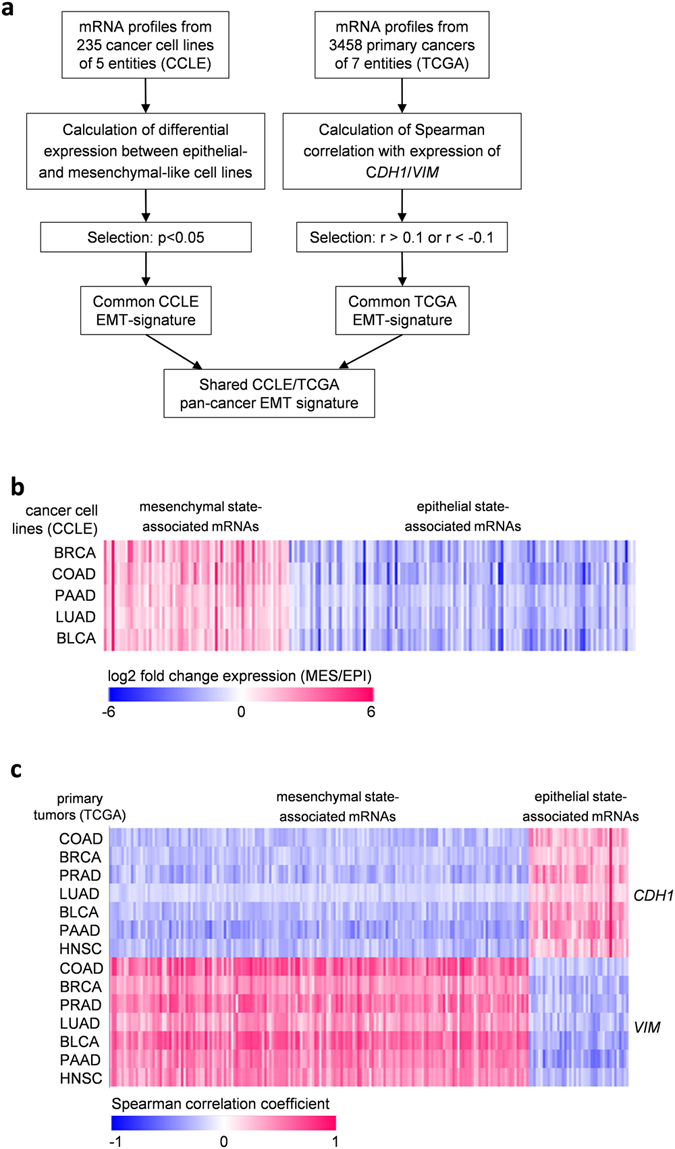
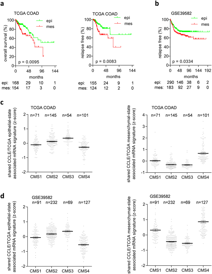
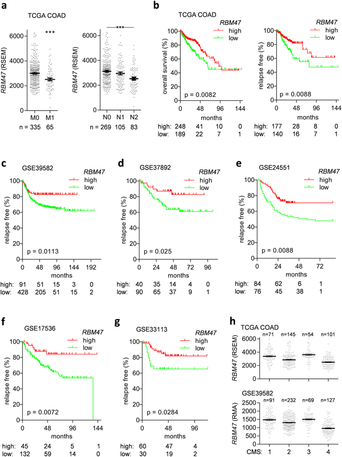
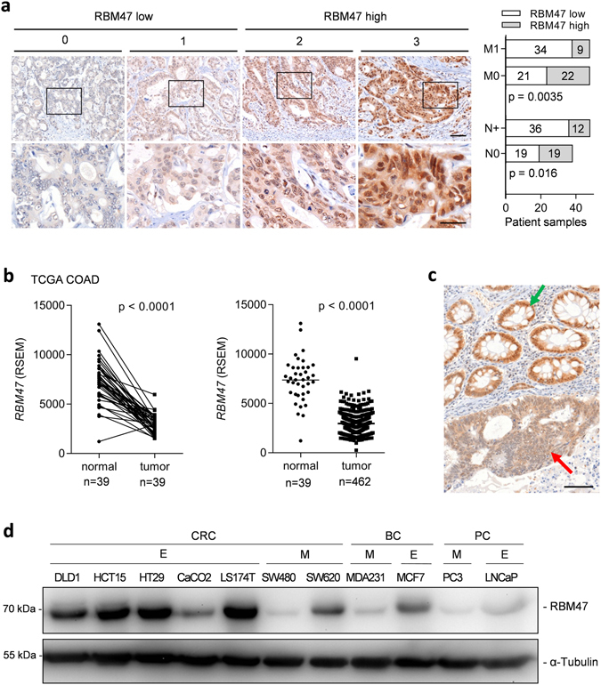
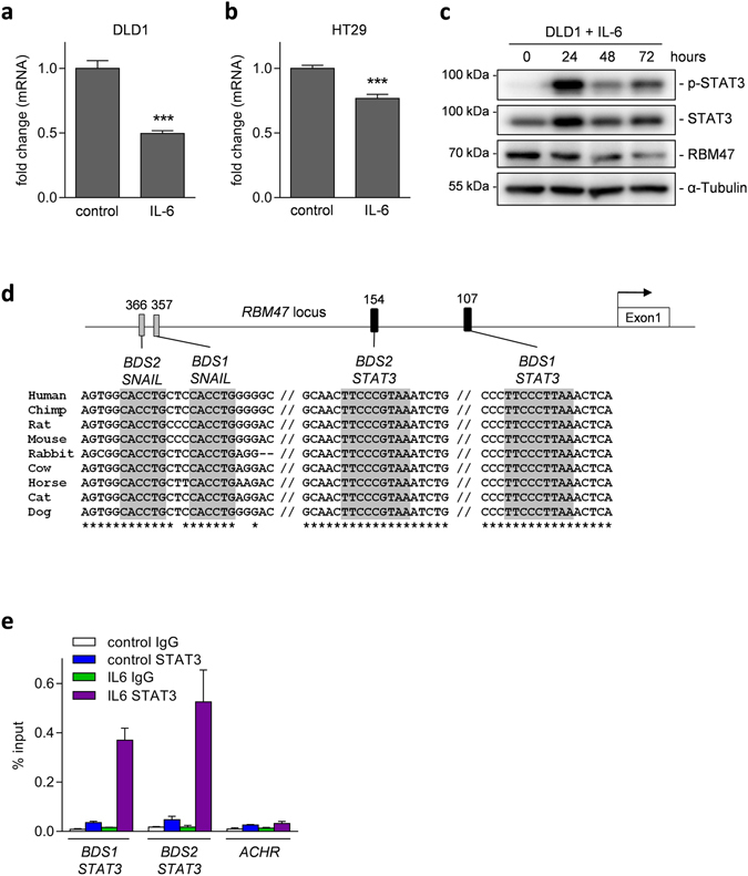
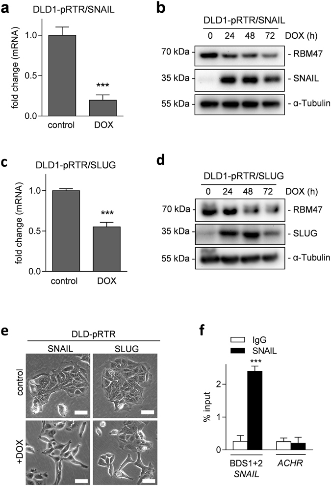
Epithelial-mesenchymal transition (EMT) plays an important role in tumor invasion and metastasis. A comprehensive, bioinformatics analysis of CCLE and TCGA datasets of seven tumor types allowed us to identify a novel pan-cancer EMT-associated gene expression signature consisting of 16 epithelial and 4 mesenchymal state-associated mRNAs. Among the identified epithelial cell state-associated factors, down-regulation of the RBM47 (RNA binding motif protein 47) mRNA displayed the most significant association with metastasis and poor survival in multiple cohorts of colorectal cancer (CRC) patients. Moreover, decreased RBM47 protein expression was associated with metastasis in a cohort of primary CRCs. RBM47 was directly suppressed during EMT induced by IL6-activated STAT3 or ectopic SNAIL and SLUG expression via conserved binding motifs of these factors within the RBM47 promoter. Moreover, RNAi-mediated down-regulation of RBM47 in CRC lines resulted in increased cell migration, invasion and metastases formation. As demonstrated by the example of RBM47, the EMT-associated signature characterized here allows to identify biomarkers for predicting clinical outcome of CRC and presumably other cancer entities. In addition, our functional analysis of RBM47 shows that the down-regulation of RBM47 during CRC progression may promote EMT and metastasis.


Conflict of interest statement

The authors declare that they have no competing interests.

Figures

Figure 1
Figure 1
Bioinformatic analysis of CCLE and TCGA datasets for EMT-associated signatures. (a) Flow chart describing the generation of the shared pan-cancer CCLE/TCGA EMT-associated mRNA signature. (b) Heatmap of 201 mRNAs that showed significant differences in expression between epithelial-like and mesenchymal-like cancer cell lines within the analysed cancer entities (log2 fold change of expression is shown; p < 0.05). (c) Heatmap showing Spearman correlation coefficients of 365 epithelial and mesenchymal state-associated mRNAs with CDH1 and VIM. Epithelial state-associated mRNAs were defined as mRNAs that positively correlate with expression of the epithelial marker CDH1 (r > 0.1) and negatively correlate with expression of the mesenchymal marker VIM (r < −0.1) in all indicated TCGA datasets. Mesenchymal state-associated mRNAs were defined as mRNAs that negatively correlate with expression of the epithelial marker CDH1 (r < −0.1) and positively correlate with expression of the mesenchymal marker VIM (r > 0.1) in all indicated TCGA datasets. COAD, colon cancer; BRCA, breast cancer; LUAD, lung cancer; HNSC, head&neck squamous cel cancer; PRAD, prostate adenocarcinomas; BLCA, bladder cancer; PAAD, pancreatic adenocarcinomas.
Figure 2
Figure 2
EMT-associated mRNA signatures are associated with survival in colon cancer patients. (a,b) Associations of epithelial state (epi) and mesenchymal state (mes) associated groups of samples with survival in TCGA COAD (a) and the GSE39582 datasets (b) (epi and mes groups of samples were defined by K-means clustering based on expression of the shared CCLE/TCGA epithelial state- or mesenchymal state-associated mRNAs. (c,d) Association of the shared TCGA/CCLE epithelial state- and mesenchymal state-associated mRNA signatures with the consensus molecular subtypes (CMS) of colorectal cancer in TCGA (c) and GSE39582 datasets (d).
Figure 3
Figure 3
Low expression of RBM47 mRNA is associated with metastasis, nodal status, and poor survival in CRC patients. (a) Association of RBM47 mRNA expression with metastasis (right) and nodal status (left) in the TCGA COAD dataset. (b) Association of RBM47 mRNA expression with overall and relapse free survival in the TCGA COAD dataset. (cg) Associations of RBM47 mRNA expression with relapse free survival in indicated CRC dataset. (h) Association of RBM47 mRNA expression with the consensus molecular subtypes (CMS) of colorectal cancer in TCGA and GSE39582 datasets.
Figure 4
Figure 4
Low expression of RBM47 protein is associated with metastasis and positive nodal status in CRC patients (a) Association of RBM47 protein expression with liver metastasis (M1) and nodal metastasis (N+) in the M0/M1 patient collection (N = 86). Left panel: representative IHC staining results of patient samples with staining scores. Right panel: quantification of IHC staining and association with metastasis (M0 – no metastases present, M1 – liver metastases present) and nodal status (N0 – no tumor cells present in lymph nodes, N+ - tumor cells present in lymph nodes). The scale bar in low magnification images (upper part) represents 100 μm and the bar in high magnification images (lower part) represents 50 μm. (b) Expression of RBM47 mRNA in normal colonic tissue and colon tumors in the TCGA COAD dataset. In the left panel expression of RBM47 in paired samples from the same patient are shown and in the right panel all samples are shown. (c) Exemplary IHC results showing expression of RBM47 protein in a colon cancer (red arrow) and adjacent normal colonic mucosa (green arrow). The scale bar represents 100 μm. (d) Immunoblot analysis of RBM47 expression in a cell line panel consisting of epithelial-like (E) and mesenchymal-like (M) colorectal (CRC), breast (BC), and prostate (PC) cancer cell lines.
Figure 5
Figure 5
Direct suppression of RBM47 by IL-6 activated STAT3. (a and b) Expression of RBM47 mRNA in (a) DLD1 or (b) HT29 CRC cells after treatment with IL-6 (20 ng/ml) or vehicle for 72 hours. (c) Western blot analysis of indicated proteins in DLD1 cells after exposure to IL-6 (20 ng/ml) for the indicated periods. (d) Map of the human RBM47 promoter region with the indicated conserved STAT3 and SNAIL binding sites. Filled rectangles represent the binding sites. TF binding sequence motifs are indicated by grey shadowing. Their conservation between species is indicated by asterisks. The arrow indicates the TSS, // represent additional, not shown sequences between the BDS. (e) qChIP analysis of STAT3 occupancy at the RBM47 promoter and, as a control, the acetylcholine receptor (ACHR) locus in DLD1 cells treated with vehicle or IL-6 for 20 minutes. In a,b, and e mean values ± SD (n = 3) are provided with *P < 0.05; **P < 0.01; ***P < 0.001.
Figure 6
Figure 6
Direct suppression of RBM47 by the EMT-TFs SNAIL and SLUG. (a) qPCR analysis of RBM47 mRNA and (b) Western blot analysis of the indicated proteins in DLD1 cell pools harboring pRTR/SNAIL plasmids. In (a) cells were treated with DOX or vehicle for 72 hours, in (b) with DOX for the indicated periods. (c and d) as in (a) and (b) but using DLD1 cell pools harboring pRTR/SLUG plasmids. (e) Phase contrast microscopy of the DLD-pRTR/SNAIL and DLD-pRTR/SLUG cell lines treated with vehicle or DOX for 48 hours. Scale bars represent 50 μm. (f) qChIP analysis of SNAIL occupancy at the RBM47 promoter and, as a control, the acetylcholine receptor (ACHR) locus in SW480 cells. In a, c, and f mean values ± SD (n = 3) are provided. *P < 0.05; **P < 0.01; ***P < 0.001.
Figure 7
Figure 7
Inhibition of RBM47 induces EMT, cell migration and invasion. (a) Phase contrast microscopy of the indicated CRC cell lines transfected with control or RBM47-specific siRNA. Scale bars represent 100 μm. (b) Western blot analysis of the indicated proteins in CRC cell lines transfected with control or RBM47-specific siRNA for 72 h. (c) qPCR analysis of the indicated mRNAs in the indicated CRC cell lines transfected with control or RBM47-specific siRNA for 72 h. (d) Wound healing assay of the indicated CRC cell lines transfected with control or RBM47-specific siRNA 48 hours before a scratch was generated. (Upper panels) Representative photographs of the initial wound area and the same area 24 hours later. (Lower panels) Quantification of wound closures: The width of scratches in 2 independent wells was analyzed for each state. Results represent the average (%) of wound closure. (e) Relative invasion of indicated CRC cells in matrigel-coated Boyden chambers transfected with control or RBM47-specific siRNA for 48 hours. In c, d, and e mean values ± SD (n = 3) are provided. *P < 0.05; **P < 0.01; *** P < 0.001.
Figure 8
Figure 8
Inhibition of RBM47 induces metastasis. Formation of lung metastases after tail-vein injection of DLD-1–Luc2 cells transfected with control or RBM47-specific siRNA. (a) Representative images of luciferase signals after D-luciferin injection at the indicated time points after xenografting. (b) Weekly measurements of total photon flux. Results are the mean ± SD (n = 5). **P < 0.01. (c) Representative lungs 8 weeks after tail vein injection. (d) H&E staining of lung tissue. Scale bars represent 500 μm. (e) Number of metastatic nodules in lungs 8 weeks after tail vein injection of the indicated DLD1 cells into mice (n = 5). (f) Model of regulation of RBM47 expression and its downstream targets, which presumably mediate its effects on EMT and cancer progression.

Similar articles

Cited by

References

    1. Turajlic S, Swanton C. Metastasis as an evolutionary process. Science. 2016;352:169–175. doi: 10.1126/science.aaf2784. - DOI - PubMed
    1. Gupta GP, Massague J. Cancer metastasis: building a framework. Cell. 2006;127:679–695. doi: 10.1016/j.cell.2006.11.001. - DOI - PubMed
    1. Nguyen DX, Bos PD, Massague J. Metastasis: from dissemination to organ-specific colonization. Nat Rev Cancer. 2009;9:274–284. doi: 10.1038/nrc2622. - DOI - PubMed
    1. Kalluri R, Weinberg RA. The basics of epithelial-mesenchymal transition. J Clin Invest. 2009;119:1420–1428. doi: 10.1172/JCI39104. - DOI - PMC - PubMed
    1. Thiery JP, Acloque H, Huang RY, Nieto MA. Epithelial-mesenchymal transitions in development and disease. Cell. 2009;139:871–890. doi: 10.1016/j.cell.2009.11.007. - DOI - PubMed

Publication types

MeSH terms