. 2017 Jul 5;7(1):4657.
doi: 10.1038/s41598-017-04730-5.

Basal hypersecretion of glucagon and insulin from palmitate-exposed human islets depends on FFAR1 but not decreased somatostatin secretion

Affiliations

Basal hypersecretion of glucagon and insulin from palmitate-exposed human islets depends on FFAR1 but not decreased somatostatin secretion

H Kristinsson et al. Sci Rep. .

Abstract

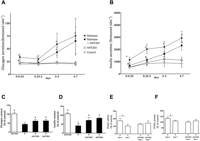
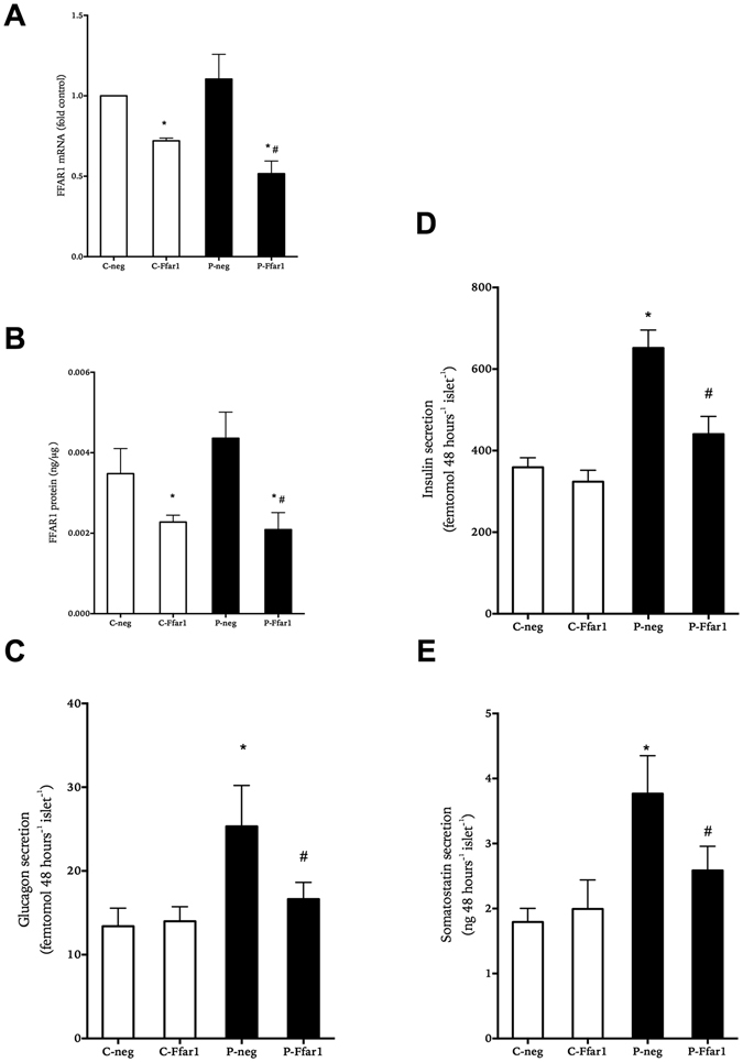
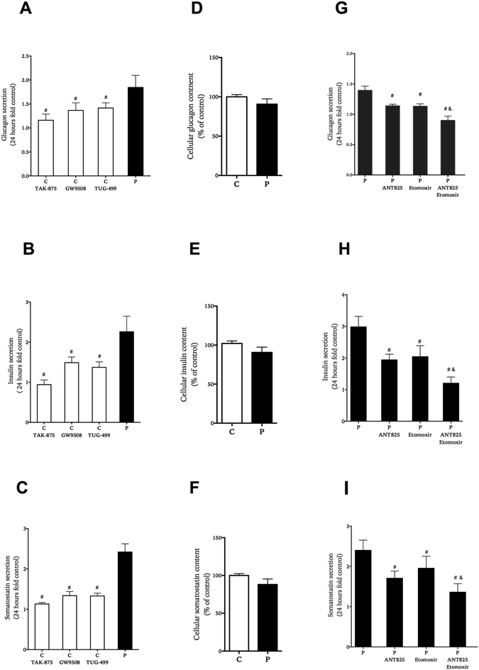
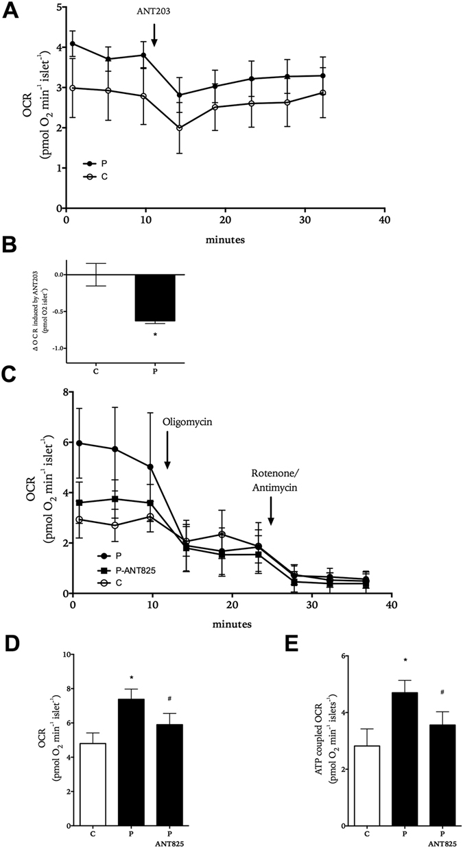
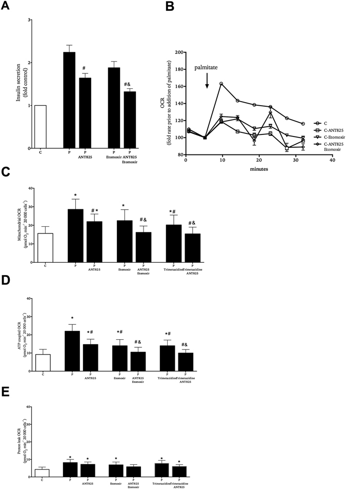
In obesity fasting levels of both glucagon and insulin are elevated. In these subjects fasting levels of the free fatty acid palmitate are raised. We have demonstrated that palmitate enhances glucose-stimulated insulin secretion from isolated human islets via free fatty acid receptor 1 (FFAR1/GPR40). Since FFAR1 is also present on glucagon-secreting alpha-cells, we hypothesized that palmitate simultaneously stimulates secretion of glucagon and insulin at fasting glucose concentrations. In addition, we hypothesized that concomitant hypersecretion of glucagon and insulin was also contributed by reduced somatostatin secretion. We found basal glucagon, insulin and somatostatin secretion and respiration from human islets, to be enhanced during palmitate treatment at normoglycemia. Secretion of all hormones and mitochondrial respiration were lowered when FFAR1 or fatty acid β-oxidation was inhibited. The findings were confirmed in the human beta-cell line EndoC-βH1. We conclude that fatty acids enhance both glucagon and insulin secretion at fasting glucose concentrations and that FFAR1 and enhanced mitochondrial metabolism but not lowered somatostatin secretion are crucial in this effect. The ability of chronically elevated palmitate levels to simultaneously increase basal secretion of glucagon and insulin positions elevated levels of fatty acids as potential triggering factors for the development of obesity and impaired glucose control.


Conflict of interest statement

H.K., E.S., H.M. and P.B. have nothing to disclose. S.O.G. and D.M.S. are employed by AstraZeneca.

Figures

Figure 1
Figure 1
Effect of palmitate on accumulated glucagon (panel A) and insulin secretion (panel B) from isolated human islets cultured for 0.25 to 7 days at 5.6 mM glucose in the absence (white symbols) or presence (black symbols) of palmitate with and without FFAR1 antagonist ANT203. Cellular content of glucagon (panel C) and insulin (panel D) at the end of culture in palmitate-treated islets with and without FFAR1 antagonists ANT203 and ANT825 presented as % of the hormone content in islets receiving control treatment. Levels of FFAR1 mRNA (panel E) and protein (panel F) in palmitate-treated islets with and without FFAR1 antagonist ANT203. Transcript and protein levels presented as % of levels in islets receiving control treatment. Results show mean ± SEM from n = 3–5 experiments donor experiments. *P < 0.05 vs. control and #p < 0.05 vs. palmitate.
Figure 2
Figure 2
Effect of decreasing FFAR1 expression on accumulated hormone secretion from human islets cultured at 5.6 mM glucose in the absence (white bars) or presence of 0.5 mM palmitate (black bars) for 1 day. Relative FFAR1 mRNA levels (panel A) and total FFAR1 protein (panel B) at the end of culture as compared to non-mammalian target shRNA (neg) or shRNA targeting Ffar1 (Ffar1) treatment. Accumulated secretion of glucagon (panel C), insulin (panel D) and somatostatin (panel E) is shown. Results show mean ± SEM and are from n = 3–5 donor experiments. *P < 0.05 vs. control-neg, #p < 0.05 vs. palmitate-neg.
Figure 3
Figure 3
Effect of FFAR1 and fatty acid β-oxidation on basal glucagon (panels A,G), insulin (panels B,H) and somatostatin (panels C,I) secretion and content (panels D-F,F). Human islets were cultured for 1 day with or with out FFAR1 agonists; TAK-875, GW9508, TUG-499 in comparison to palmitate (black bars) (panel A–C), and with palmitate with and without ANT825 and/or etomoxir (panel G–I). Results show mean ± SEM and are from n = 5 donor experiments. #P < 0.05 vs. palmitate, and & vs. palmitate + etomoxir.
Figure 4
Figure 4
Role of FFAR1 in the effect of palmitate on human islet respiration. OCR recordings from human islets exposed to 5.6 mM glucose (white symbols) and 0.5 mM palmitate (black symbols) and sequential addition of ANT203 (panel A). Difference in OCR 20 minutes prior and post addition of ANT203 (panel B). OCR recordings from human islets exposed for 2 hours to 5.6 mM glucose (white symbols), 0.5 mM palmitate (black circles, black bars) or palmitate with ANT825 (black triangles, black bars) and the sequential addition of oligomycin and rotenone/antimycin (panel C). Mitochondrial respiration (panel D) and ATP-coupled respiration (panel E) are shown. Results show mean ± SD in (panels A−C) (N = 10) and mean ± SEM in (panels D and E) (n = 5–7 donor experiments). *P < 0.05 vs. control and #p < 0.05 vs. palmitate.
Figure 5
Figure 5
Role of FFAR1 and fatty acid β-oxidation in the effect of palmitate on EndoC-βH1 insulin secretion and respiration. Cells were exposed to 5.6 mM glucose in the absence (white symbols) or presence (black symbols) of palmitate with and without ANT825 and/or etomoxir, trimetazidine. Insulin secretion (panel A). Representative kinetic OCR recordings from cells acutely exposed to palmitate with and without ANT825 and/or etomoxir (panel B). Mitochondrial OCR (panel C), ATP-coupled OCR (panel D) and proton leak OCR (panel E). Results show mean ± SEM (panels A,C-E,) (n = 5), and mean ± SD (panel B) (N = 6). *P < 0.05 vs. control, #P < 0.05 vs. palmitate alone and &P < 0.05 vs. corresponding P-etomoxir/trimetazidine.

References

    1. Manell H, et al. Altered Plasma Levels of Glucagon, GLP-1 and Glicentin During OGTT in Adolescents With Obesity and Type 2 Diabetes. The Journal of clinical endocrinology and metabolism. 2016;101:1181–1189. doi: 10.1210/jc.2015-3885. - DOI - PubMed
    1. D’Alessio D. The role of dysregulated glucagon secretion in type 2 diabetes. Diabetes, obesity & metabolism. 2011;13(Suppl 1):126–132. doi: 10.1111/j.1463-1326.2011.01449.x. - DOI - PubMed
    1. Erdmann J, et al. Disturbances of basal and postprandial insulin secretion and clearance in obese patients with type 2 diabetes mellitus. Hormone and metabolic research = Hormon- und Stoffwechselforschung = Hormones et metabolisme. 2012;44:60–69. - PubMed
    1. Kozawa J, et al. Early postprandial glucagon surge affects postprandial glucose levels in obese and non-obese patients with type 2 diabetes. Endocr J. 2013;60:813–818. doi: 10.1507/endocrj.EJ13-0018. - DOI - PubMed
    1. Quesada I, Tuduri E, Ripoll C, Nadal A. Physiology of the pancreatic alpha-cell and glucagon secretion: role in glucose homeostasis and diabetes. The Journal of endocrinology. 2008;199:5–19. doi: 10.1677/JOE-08-0290. - DOI - PubMed

Publication types

MeSH terms