

18F-Fluorodeoxyglucose Positron Emission Tomography/Computed Tomography Findings of Pancreatic Hemangiopericytoma
- PMID: 28680212
- PMCID: PMC5482024
- DOI: 10.4103/ijnm.IJNM_24_17
18F-Fluorodeoxyglucose Positron Emission Tomography/Computed Tomography Findings of Pancreatic Hemangiopericytoma
Abstract
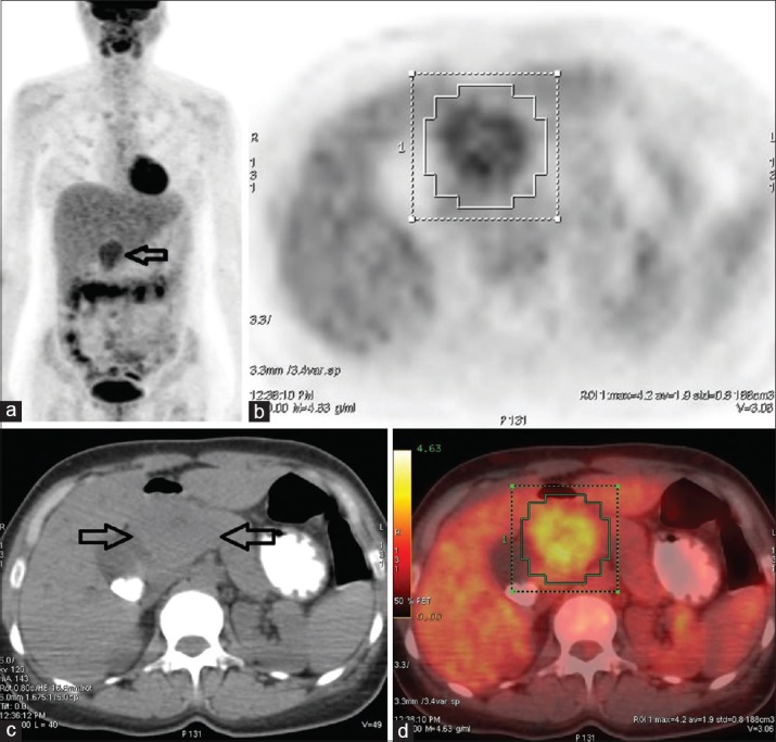
A 42-year-old woman with a large pancreatic tumor had undergone 18F-fluorodeoxyglucose positron emission tomography/computed tomography (FDG PET/CT) imaging. Moderate metabolic activity was detected on the head of the pancreas. The tumor was resected and it was histopathologically diagnosed as malign pancreatic hemangiopericytoma (HPC). HPC is a rare soft tissue sarcoma. The tumor is considered aggressive with high rates of local recurrence and metastasis regardless the localization. Herein, we present the imaging characteristics of HPC with 18F-FDG PET/CT.
Keywords: 18F-fluorodeoxyglucose positron emission tomography/computed tomography; hemangiopericytoma; pancreas.
Conflict of interest statement
There are no conflicts of interest.
Figures

References
-
- Pitluk HC, Conn J., Jr Hemangiopericytoma. Literature review and clinical presentations. Am J Surg. 1979;137:413–6. - PubMed
-
- Spitz FR, Bouvet M, Pisters PW, Pollock RE, Feig BW. Hemangiopericytoma: A 20-year single-institution experience. Ann Surg Oncol. 1998;5:350–5. - PubMed
-
- Bardaxogou E, Manganas D, Landen S, Ramée MP, Chareton B, Maddern GJ, et al. Hemangiopericytoma of the pancreas: Report of a case and review of the literature. Hepatogastroenterology. 1995;42:172–4. - PubMed
Publication types
LinkOut - more resources
Full Text Sources
Other Literature Sources