. 2017 Jun 21:11:119.
doi: 10.3389/fnbeh.2017.00119. eCollection 2017.

Botulinum Neurotoxin A Injected Ipsilaterally or Contralaterally into the Striatum in the Rat 6-OHDA Model of Unilateral Parkinson's Disease Differently Affects Behavior

Affiliations

Botulinum Neurotoxin A Injected Ipsilaterally or Contralaterally into the Striatum in the Rat 6-OHDA Model of Unilateral Parkinson's Disease Differently Affects Behavior

Veronica A Antipova et al. Front Behav Neurosci. .

Abstract

Parkinson's disease (PD) is one of the most frequent neurodegenerative disorders. The loss of dopaminergic neurons in the substantia nigra leads to a disinhibition of cholinergic interneurons in the striatum. Pharmacotherapeutical strategies of PD-related hypercholinism have numerous adverse side effects. We previously showed that ipsilateral intrastriatal injections of 1 ng in unilaterally 6-hydroxydopamine (6-OHDA)-lesioned rats inhibit apomorphine-induced rotation behavior significantly up to 6 months. In this study, we extended the behavioral testing of ipsilateral botulinum neurotoxin A (BoNT-A)-injection and additionally investigated the impact of intrastriatal BoNT-A-injections contralateral to the 6-OHDA-lesioned hemisphere on the basal ganglia circuity and motor functions. We hypothesized that the interhemispheric differences of acetylcholine (ACh) concentration seen in unilateral hemi-PD should be differentially and temporally influenced by the ipsilateral or contralateral injection of BoNT-A. Hemi-PD rats were injected with 1 ng BoNT-A or vehicle substance into either the ipsilateral or contralateral striatum 6 weeks after 6-OHDA-lesion and various behaviors were tested. In hemi-PD rats intrastriatal ipsilateral BoNT-A-injections significantly reduced apomorphine-induced rotations and increased amphetamine-induced rotations, but showed no significant improvement of forelimb usage and akinesia, lateralized sensorimotor integration and also no effect on spontaneous locomotor activity. However, intrastriatal BoNT-A-injections contralateral to the lesion led to a significant increase of the apomorphine-induced turning rate only 2 weeks after the treatment. The apomorphine-induced rotation rate decreases thereafter to a value below the initial rotation rate. Amphetamine-induced rotations were not significantly changed after BoNT-A-application in comparison to sham-treated animals. Forelimb usage was temporally improved by contralateral BoNT-A-injection at 2 weeks after BoNT-A. Akinesia and lateralized sensorimotor integration were also improved, but contralateral BoNT-A-injection had no significant effect on spontaneous locomotor activity. These long-ranging and different effects suggest that intrastriatally applied BoNT-A acts not only as an inhibitor of ACh release but also has long-lasting impact on transmitter expression and thereby on the basal ganglia circuitry. Evaluation of changes of transmitter receptors is subject of ongoing studies of our group.

Keywords: Wistar rats; behavior; botulinum toxins; hemiparkinsonism; striatum.


Figures
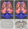
Figure 1
Figure 1
Dorsal view of reconstructed rat brains. The location of the right side 6-hydroxydopamine (6-OHDA)-injection site in the medial forebrain bundle (MFB; white oval) anterior to the substantia nigra pars compacta (SNC) and the two injection sites of botulinum neurotoxin A (BoNT-A) or vehicle (yellow spots) in the caudate putamen (CPu) ipsilateral (A) and contralateral (B) to 6-OHDA are indicated. Adjacent 20 μm coronal sections of a rat brain stained for cell bodies (C) according to Merker or for myelin (D) according to Gallyas depict the needle tract (arrow) and the needle tip (asterisk) of a BoNT-A-injection ipsilateral to the 6-OHDA-application (9 months survival). Scale bar applies to (C,D): 2 mm.
Figure 2
Figure 2
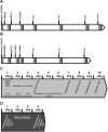
Time points of lesions and behavioral tests in rats BoNT-A or sham-injected (A) ipsilateral or (B) contralateral to 6-OHDA. Light gray rectangles symbolize batteries of spontaneous behavior tests performed subsequently: open field (OF) test, corridor task, stepping and cylinder tests. Dark gray rectangles symbolize amphetamine-induced rotation test followed by an apomorphine-induced rotation test 3 days later. (C) The battery of spontaneous behavior tests were performed as follows: each behavior test series lasted 10 days. At the first day rats were tested for 10 min in the OF arena. Thereafter, rats were food restricted and adapted 2 days rats to the corridor task apparatus for 10 min each. One day later the final corridor test was carried out for 5 min. At the following 3 days rats were handled by the experimentator for 5 min each day. On the following 3 days rats underwent the stepping test twice a day. On the 10th day the forepaw usage was evaluated by the cylinder test. (D) Finally, drug-induced rotation tests were performed. First amphetamine rotation test was done for 60 min. Three days later rats were tested for apomorphine-induced rotations for 40 min.
Figure 3
Figure 3
Apomorphine-induced rotations in hemi-Parkinson’s disease (PD) rats treated with intrastriatal BoNT-A- or vehicle-applied (A) ipsilaterally or (B) contralaterally. Ipsilateral BoNT-A-injections significantly decreased anti-clockwise rotations 4 weeks and 3 months compared to sham injection. Contralateral BoNT-A-injection caused a significant increase of the turning rate only 2 weeks after surgery. Asterisks indicate significant differences compared to the sham group (*P < 0.05). Hashtags indicate significant differences compared to values before BoNT-A-injection (##P < 0.01). Paragraphs indicate significant differences compared to the previous value after BoNT-A-injection (§§§P < 0.001). Data are represented as mean ± SEM.
Figure 4
Figure 4
Amphetamine-induced rotations in hemi-PD rats treated with intrastriatal BoNT-A-or vehicle-applied (A) ipsilaterally or (B) contralaterally. Ipsilateral BoNT-A-injection caused significantly increased rotations 4 weeks to 6 months after BoNT-A, while sham-injected rats did not change rotational behavior. Contralateral BoNT-A-injection in hemi-PD rats caused no significant change of rotational behavior compared to the sham group. Asterisks indicate significant differences compared to the sham group (*P < 0.05). Hashtags indicate significant differences compared to values before BoNT-A-injection (##P < 0.01, ###P < 0.001). Data are represented as mean ± SEM.
Figure 5
Figure 5
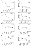
Stepping test in right side hemi-PD rats treated with intrastriatal BoNT-A- or vehicle-applied (A,C,E,G) ipsilaterally or (B,D,F,H) contralaterally. In unlesioned rats 9–12 adjusting steps for the left (A–D) and right (E–H) forelimbs in forehand and backhand directions were seen. In hemi-PD rats the use of the left forepaw was impaired in both the forehand (A,B) and backhand (C,D) directions. Neither ipsilateral BoNT-A nor sham injection changed the impairments of left paw steps in hemi-PD rats (A,C). The stepping of the right forelimb in hemi-PD rats in both the forehand (E) and backhand (G) directions were generally not affected by ipsilateral BoNT-A or sham injection. Contralateral BoNT-A-injected hemi-PD rats significantly improved left paw forehand steps from 2 weeks to 3 months after BoNT-A (B), and backhand steps from 2 weeks until 9 months after BoNT-A (D). Right forepaw adjusting steps significantly increased from 2 weeks up to 9 months after BoNT-A-application as well in forehand (F) as in backhand (H) directions compared to sham-injected rats. Asterisks indicate significant differences compared to the sham group (*P < 0.05, **P < 0.01, ***P < 0.001). Data are represented as mean ± SEM.
Figure 6
Figure 6
Cylinder test in right side hemi-PD rats treated with intrastriatal BoNT-A- or vehicle-applied (A) ipsilaterally or (B) contralaterally. Hemi-PD rats exhibited a significantly reduced use of the left forelimb resulting in left/right ratio of about 0.5 (A,B). Neither ipsilateral BoNT-A nor sham injection showed any significant improvement. Contralateral intrastriatal injection of BoNT-A only led at 2 weeks after BoNT-A to a significant readjustment of the right and left forepaw usage. Hashtags indicate significant differences compared to values before BoNT-A-injection (###P < 0.001). Asterisks indicate significant differences compared to the sham group (***P < 0.001). Data are represented as mean ± SEM.
Figure 7
Figure 7
OF test in right side hemi-PD rats treated with intrastriatal BoNT-A- or vehicle-applied (A,C) ipsilaterally or (B,D) contralaterally. Rats of both BoNT-A- and sham-injected groups showed decreased locomotor activity compared to prelesion results (B). Neither ipsilateral BoNT-A nor sham injection showed any significant changes in total running distance (A) or the ratio of center distance to total distance (C). The same is true for both contralaterally injected groups (B,D). Data are represented as mean ± SEM.
Figure 8
Figure 8
Corridor task in hemi-PD rats treated with intrastriatal BoNT-A- or vehicle-applied (A) ipsilaterally or (B) contralaterally. Preoperative screening showed that all animals equally retrieved pellets from either left or right sides (A,B). Right side hemi-PD rats significantly neglected of the left corridor side. Ipsilateral BoNT-A as well as sham injections did not improve contralateral sensorimotor integration in hemi-PD rats. Contralateral BoNT-A-injection reversed this bias in hemi-PD rats 2 weeks to 9 months. Significant contralateral retrievals of about 40% were seen. Left side sham-injected rats did not improve in this task over time. Asterisks indicate significant differences compared to the sham group (**P < 0.01). Hashtags indicate significant differences compared to values before BoNT-A-injection (###P < 0.001). Data are represented as mean ± SEM.

References

    1. Abedi P. M., Delaville C., De Deurwaerdère P., Benjelloun W., Benazzouz A. (2013). Intrapallidal administration of 6-hydroxydopamine mimics in large part the electrophysiological and behavioral consequences of major dopamine depletion in the rat. Neuroscience 236, 289–297. 10.1016/j.neuroscience.2013.01.043 - DOI - PubMed
    1. Alam M., Schmidt W. J. (2002). Rotenone destroys dopaminergic neurons and induces parkinsonian symptoms in rats. Behav. Brain Res. 136, 317–324. 10.1016/s0166-4328(02)00180-8 - DOI - PubMed
    1. Andringa G., Van Oosten R. V., Unger W., Hafmans T. G. M., Veening J., Stoof J. C., et al. . (2000). Systemic administration of the propargylamine CGP 3466B prevents behavioural and morphological deficits in rats with 6-hydroxydopamine-induced lesions in the substantia nigra. Eur. J. Neurosci. 12, 3033–3043. 10.1046/j.1460-9568.2000.00181.x - DOI - PubMed
    1. Antipova V., Hawlitschka A., Mix E., Schmitt O., Dräger D., Benecke R., et al. . (2013). Behavioral and structural effects of unilateral intrastriatal injections of botulinum neurotoxin a in the rat model of Parkinson’s disease. J. Neurosci. Res. 91, 838–847. 10.1002/jnr.23210 - DOI - PubMed
    1. Araki T., Tanji H., Kato H., Itoyama Y. (1998). Sequential changes of dopaminergic receptors in the rat brain after 6-hydroxydopamine lesions of the medial forebrain bundle. J. Neurol. Sci. 160, 121–127. 10.1016/s0022-510x(98)00248-2 - DOI - PubMed

LinkOut - more resources