. 2017 Apr 20;6(6):e1316439.
doi: 10.1080/2162402X.2017.1316439. eCollection 2017.

MYCN is an immunosuppressive oncogene dampening the expression of ligands for NK-cell-activating receptors in human high-risk neuroblastoma

Affiliations

MYCN is an immunosuppressive oncogene dampening the expression of ligands for NK-cell-activating receptors in human high-risk neuroblastoma

Elisa Brandetti et al. Oncoimmunology. .

Abstract

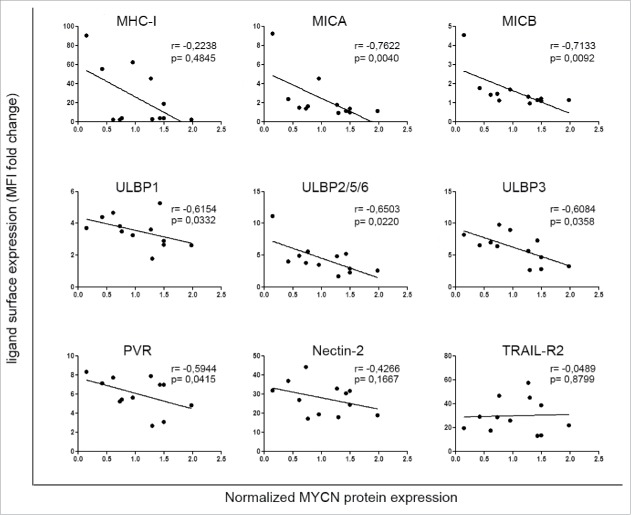
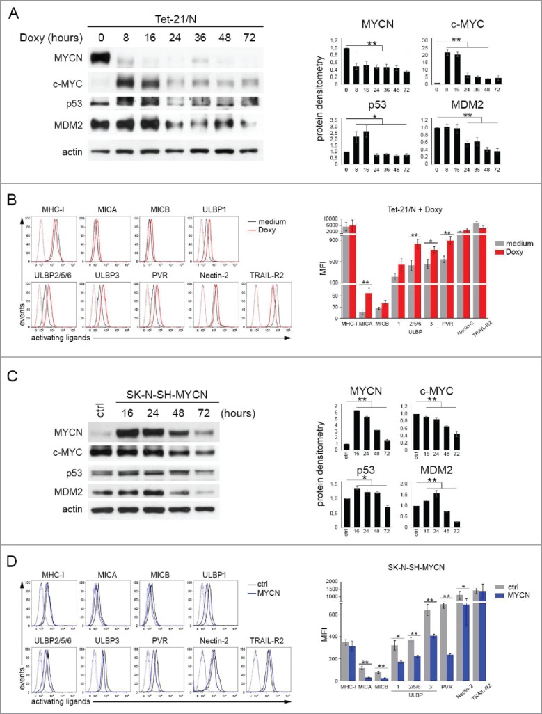
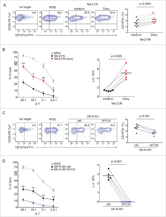
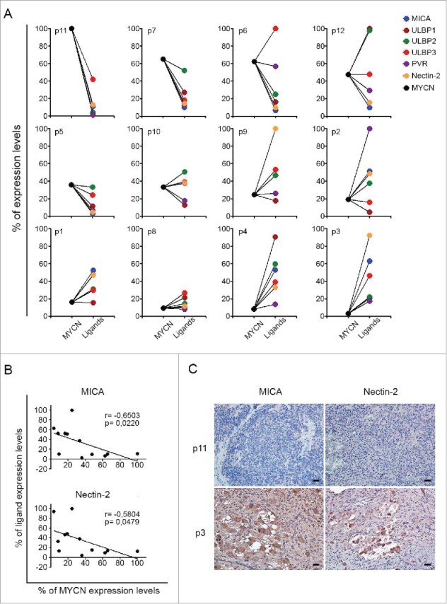
Neuroblastoma (NB) is the most common extracranial solid tumor occurring in childhood. Amplification of the MYCN oncogene is associated with poor prognosis. Downregulation on NB cells of ligands recognized by Natural Killer (NK) cell-activating receptors, involved in tumor cell recognition and lysis, may contribute to tumor progression and relapse. Here, we demonstrate that in human NB cell lines MYCN expression inversely correlates with that of ligands recognized by NKG2D and DNAM1 activating receptors in human NB cell lines. In the MYCN-inducible Tet-21/N cell line, downregulation of MYCN resulted in enhanced expression of the activating ligands MICA, ULBPs and PVR, which rendered tumor cells more susceptible to recognition and lysis mediated by NK cells. Conversely, a MYCN non-amplified NB cell line transfected with MYCN showed an opposite behavior compared with control cells. Consistent with these findings, an inverse correlation was detected between the expression of MYCN and that of ligands for NK-cell-activating receptors in 12 NB patient specimens both at mRNA and protein levels. Taken together, these results provide the first demonstration that MYCN acts as an immunosuppressive oncogene in NB cells that negatively regulates the expression of ligands for NKG2D and DNAM-1 NK-cell-activating receptors. Our study provides a clue to exploit MYCN expression levels as a biomarker to predict the efficacy of NK-cell-based immunotherapy in NB patients.

Keywords: Immunosuppressive oncogene; MYCN oncogene; NK-cell-activating receptor ligands; neuroblastoma; tumor immune escape.


Figures

Figure 1.
Figure 1.
Expression of MYCN inversely correlates with that of ligands for NK-cell-activating receptors in NB cell lines. Scatter plots showing the correlation between MYCN and the surface expression of activating ligands for NK-cell receptors in 12 NB cell lines. The means of four independent immunoblotting analyses of MYCN expression in NB cell lines are plotted against the means of 15 independent flow cytometric analyses of surface expression of the indicated activating ligands (see Table S1 for data); Spearman correlation r and p value are shown for each plot.
Figure 2.
Figure 2.
Modulation of MYCN affects the expression of ligands for NK-cell-activating receptors. (A) Representative example of immunoblot analysis of MYCN, c-MYC, p53 and MDM2 in Tet-21/N either left untreated (0) or treated with doxycycline for the indicated time (left panel). An anti-Actin Ab was used for normalization. Densitometry analysis of Actin-normalized proteins values of three independent experiments are shown (right panel). Mean ± SD; *p < 0.05, **p < 0.01. (B) Representative flow cytometric analysis of surface expression of activating ligands for NK-cell receptors in Tet-21/N either left untreated (medium, gray line) or treated with doxycycline for 16 h (Doxy, red line); dotted lines, isotype-matched negative controls (left panel). Summary of five independent flow cytometric analyses (right panel). p values, compared with untreated and Doxy-treated Tet-21/N cells (two-tailed paired Student's t-test); *p < 0.05, **p < 0.01. (C) Representative example of immunoblot analysis as in A of SK-N-SH either left transfected with empty vector (ctrl) or with piRVneoSV-MYCN (MYCN) for the indicated time (left panel). Densitometry analysis of Actin-normalized proteins values of three independent experiments are shown (right panel). Mean ± SD; *p < 0.05, **p < 0.01. (D) Representative flow cytometric analysis of surface expression of activating ligands for NK-cell receptors in SK-N-SH transfected for 48 h with control vector (ctrl, gray line) or with MYCN cDNA vector (MYCN, blue line); dotted lines, isotype-matched negative controls (left panel). Summary of five independent flow cytometric analyses (right panel). p values, compared with ctrl and MYCN cDNA vector transfected cells (two-tailed paired Student's t-test); *p < 0.05, **p < 0.01.
Figure 3.
Figure 3.
Modulation of MYCN renders NB cells differently susceptible to NK-cell-mediated lysis. (A) Degranulation of human CD3CD56+ CD45+ NK cells from healthy donors, measured as CD107a cell-surface expression following stimulation with Tet-21/N cells, either left untreated (medium) or treated with doxycycline for 16 h (Doxy). K562 cells were used as positive control. The percentage of CD107a+ NK cells is indicated. A representative experiment out of the eight performed is shown (left panel). Summary of NK-cell degranulation of cells isolated from eight healthy donors is shown (right panel). Dots, percentage of CD107a+ NK cells; horizontal bars, average values. Dashed lines connect percentage of CD107a+ NK cells from each donor cells. p values, compared with untreated and doxycycline-treated Tet-21/N cells (two-tailed paired Student's t-test). (B) Tet-21/N untreated or treated as in (A) were tested as targets for NK cells at the indicated effector:target (E:T) ratios in a standard 51Cr-release assay. One representative experiment out of the five performed is shown (left panel). p values, compared with untreated and doxycycline-treated Tet-21/N cells (two-tailed paired Student's t-test); *p < 0.05, **p < 0.01. Summary of cytotoxic assay of Tet-21/N cells treated as in A and tested as targets of NK cells isolated from five healthy donors in a standard 51Cr-release assay (right panel). Specific lysis was converted to L.U. 20%. Dots, L.U. 20% of the effector/target pairs tested; horizontal bars, average values. Dashed lines connect L.U. 20% from each donor cells obtained with the indicated target. p values, compared with untreated and doxycycline-treated Tet-21/N cells (two-tailed paired Student's t-test). (C) Degranulation assay of human NK cells, as in (A), following stimulation with SK-N-SH cells transfected for 48 h with piRVneoSV vector (ctrl) or with piRVneoSV-MYCN (MYCN). One representative experiment out of the six performed (left panel) and the summary of six experiments (right panel) are shown. (D) Cytotoxic activity of healthy donor NK cells, as in (B), against SK-N-SH cells transfected for 48 h with ctrl or MYCN cDNA vector. One representative experiment out of the five performed (left panel) and the summary of five experiments (right panel) are shown.
Figure 4.
Figure 4.
Expression of MYCN and of NK-cell-activating receptor ligands in primary NB cells. (A) qPCR analysis of MYCN and the indicated activating ligands in 12 primary NB samples. GAPDH was used for normalization. Data were normalized as percentages of the highest value obtained for each marker and plotted as percentage of expression levels. Data are representative of four experiments. (B) Scatter plots showing the correlation between MYCN and both MICA and Nectin-2 in 12 NB samples. The means of four independent qPCR analyses, expressed as percentage of expression levels, of MYCN are plotted against those of MICA and Nectin-2; Spearman correlation r and p value are shown for each plot. (C) Expression of MICA and Nectin-2 in primary NB samples by immunohistochemistry assay. MICA and Nectin-2 expressing cells are shown in brown. Nuclei are counterstained with hematoxylin (blue). Original magnification, ×20. Scale bars 30 µm.

References

    1. Maris JM. Recent advances in neuroblastoma. N Eng J Med 2010; 362:2202-11; PMID:20558371; https://doi.org/10.1056/NEJMra0804577 - DOI - PMC - PubMed
    1. Zimmerman KA, Yancopoulos GD, Collum RG, Smith RK, Kohl NE, Denis KA, Nau MM, Witte ON, Toran-Allerand D, Gee CE et al. . Differential expression of MYC family genes during murine development. Nature 1986; 319:780-3; PMID:2419762; https://doi.org/10.1038/319780a0 - DOI - PubMed
    1. Raffaghello L, Prigione I, Bocca P, Morandi F, Camoriano M, Gambini C, Wang X, Ferrone S, Pistoia V. Multiple defects of the antigen-processing machinery components in human neuroblastoma: immunotherapeutic implications. Oncogene 2005; 24:4634-44; PMID:15897905; https://doi.org/10.1038/sj.onc.1208594 - DOI - PubMed
    1. Marcus A, Gowen BG, Thompson TW, Iannello A, Ardolino M, Deng W, Wang L, Shifrin N, Raulet DH. Recognition of tumors by the innate immune system and natural killer cells. Adv Immunol 2014; 122:91-128; PMID:24507156; https://doi.org/10.1016/B978-0-12-800267-4.00003-1 - DOI - PMC - PubMed
    1. Raffaghello L, Prigione I, Airoldi I, Camoriano M, Levreri I, Gambini C, Pende D, Steinle A, Ferrone S, Pistoia V. Downregulation and/or release of NKG2D ligands as immune evasion strategy of human neuroblastoma. Neoplasia 2004; 6:558-68; PMID:15548365; https://doi.org/10.1593/neo.04316 - DOI - PMC - PubMed

Publication types

LinkOut - more resources