. 2017 Apr 18;6(6):e1317411.
doi: 10.1080/2162402X.2017.1317411. eCollection 2017.

Tumor-priming converts NK cells to memory-like NK cells

Affiliations

Tumor-priming converts NK cells to memory-like NK cells

Marina Pal et al. Oncoimmunology. .

Abstract

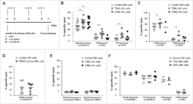
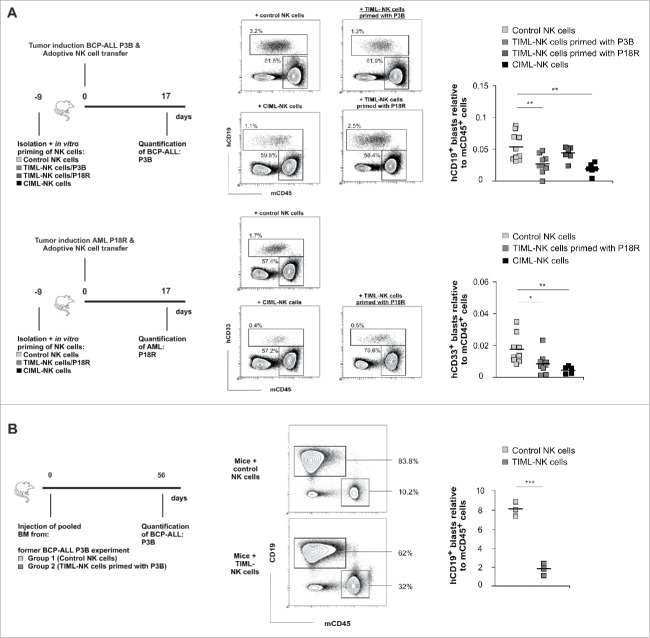
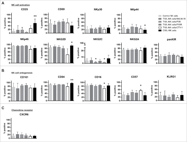
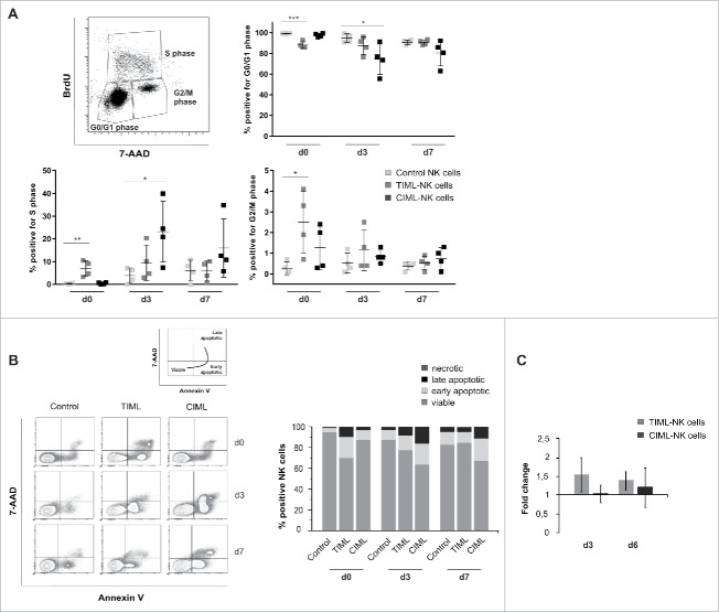
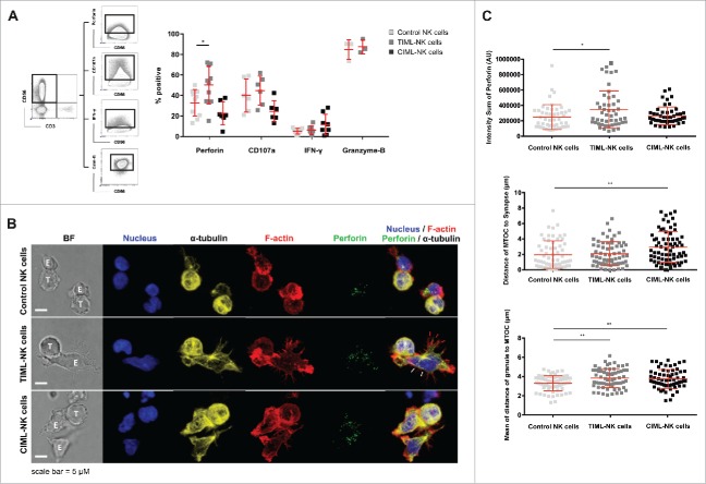
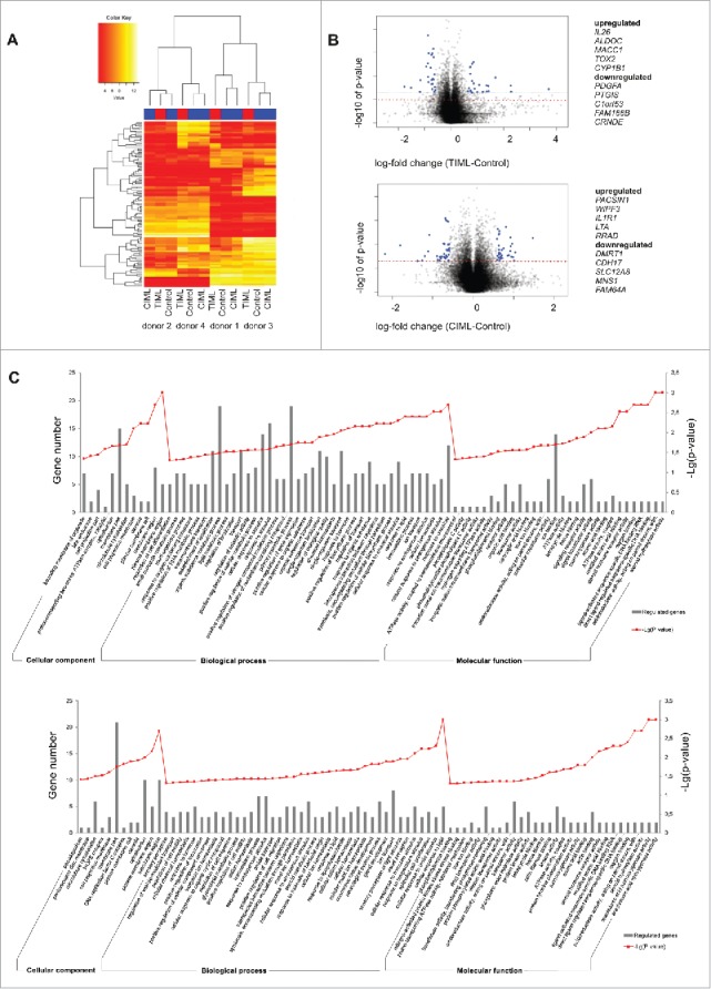
Fascinating earlier evidence suggests an intrinsic capacity of human natural killer (NK) cells to acquire adaptive immune features in the context of cytomegalovirus (CMV) infection or pro-inflammatory cytokine stimulation. Since the role of memory NK cells in cancer has so far remained elusive and adoptive NK cell transfer in relapsing pediatric acute B cell precursor leukemia (BCP-ALL) patients awaits improvement, we asked the question whether tumor-priming could promote the generation of memory NK cells with enhanced graft-vs.-leukemia (GvL) reactivity. Here, we provide substantial evidence that priming of naive human NK cells with pediatric acute B cell leukemia or acute myeloid leukemia specimens induces a functional conversion to tumor-induced memory-like (TIML)-NK cells displaying a heightened tumor-specific cytotoxicity and enhanced perforin synthesis. Cell cycles analyses reveal that tumor-priming sustainably alters the balance between NK cell activation and apoptosis in favor of survival. In addition, gene expression patterns differ between TIML- and cytokine-induced memory-like (CIML)-NK cells with the magnitude of regulated genes being distinctly higher in TIML-NK cells. As such, the tumor-induced conversion of NK cells triggers the emergence of a so far unacknowledged NK cell differentiation stage that might promote GvL effects in the context of adoptive cell transfer.

Keywords: Acute B cell precursor leukemia; adoptive cell transfer; children; natural killer cells.


Figures

Figure 1.
Figure 1.
Tumor-priming induces TIML-NK cells to elicit a superior, tumor-restricted functionality against pediatric BCP-ALL and AML. (A) Experimental layout for in vitro generation of TIML-NK cells. Freshly isolated NK cells were primed on d-1 with different irradiated tumor specimens, irradiated PBMCs or with a mixture of 10 ng/mL IL12 and 50 ng/mL IL18. All NK cell preparations were cultured in medium supplemented with low dose (100 IU/mL) IL2 and low dose (1 ng/mL) IL15. Cytotoxicity was tested on d7. (B) BCP-ALL-primed TIML-NK cells exhibit heightened anti-tumor functionality toward pediatric BCP-ALL. In vitro cytotoxicity assays on d7. Unprimed (“control NK cells”), BCP-ALL (NALM-16-, P3B- or P31G)-primed (“TIML-NK cells”) and IL12/IL18-primed (“CIML-NK cells”) NK cells were used as effectors and the identical tumor specimen was used as a target for re-stimulation on d7. Data represent 10 (NALM-16 priming/re-stimulation), 7 (P3B-priming/re-stimulation) or 5 (P31G-priming/re-stimulation) different donors (E:T ratio 3:1 in NALM-16 and P3B experiments, E:T ratio 9:1 in P31G experiments). (C) AML-primed TIML-NK cells exhibit heightened anti-tumor functionality toward the identical pediatric AML. In vitro cytotoxicity assays on d7. Unprimed, AML (P18R- or P84D)-primed and IL12/IL18-primed NK cells were used as effectors and the identical tumor specimen was used as a target for re-stimulation on d7. Data represent 5 (P18R priming/re-stimulation) or 3 (P84D-priming/re-stimulation) different donors (E:T ratio 3:1 in all experiments). (D) Priming-induced NK cell conversion requires exposure to malignant cells. NK cells from donors depicted in Fig. 1B (NALM-16-priming) were primed with irradiated allogeneic PBMCs at a ratio of 1:3. In vitro cytotoxicity assays performed on d7 with control or PBMC-primed NK cells as effectors and NALM-16 cells as targets. Results represent data from six different NK cell-donors primed with 5 different PBMC specimens (E:T ratio 1:1). (E) NALM-16-primed TIML-NK cells do not exert cytotoxicity toward non-malignant PBMCs. In vitro cytotoxicity assays were performed on d7 with NALM-16-primed NK cells as effectors and autologous or allogeneic PBMCs as targets. Data represent three different donors (E:T ratio 1:1). (F) TIML-NK cells show heightened cytotoxicity only toward the original priming tumor entity. Unprimed, NALM-16-, P31G-, P3B- or P18R-primed and IL12/IL18-primed NK cells were used as effectors; as indicated other tumor specimens were used targets for re-stimulation on d7 to test functional TIML-NK cell specificity. Note, that the donors shown in Fig. 1F are identical to the respective donors tested in Fig. 1B and C, i.e., the efficacy of the priming effect was documented for every donor shown in Fig. 1F. Data represent 3 (NALM-16 priming/Kasumi-1 re-stimulation), 5 (P31G priming/NALM-16 re-stimulation), 3 (P3B priming/P18R re-stimulation) or 4 (P18R priming/P3B re-stimulation) different donors. E:T ratio 3:1 (all experiments). All experiments were performed in triplicates. **p < 0.01, ***p < 0.001.
Figure 2.
Figure 2.
TIML-NK cells confer anti-leukemic activity toward pediatric BCP-ALL and AML in vivo. (A) Experimental layouts for in vivo experiments. Blasts of the donors P3B (BCP-ALL) or P18R (AML) were injected on d0 into NSG mice followed by a second injection of the varying NK cell preparations as indicated several hours later on d0. Leukemic burden was quantified in the bone marrow 17 d later. Exemplified gating strategy for identification of the leukemic load shown as human CD19+ blasts (for the BCP-ALL P3B) or human CD33+ (for the AML P18R) relative to murine CD45+ cells, respectively. Data from BCP-ALL (P3B)-tumor induction represent n = 10 mice injected with control NK cells (three different donors), n = 9 mice injected with TIML-NK cells (three different donors) primed with P3B blasts, n = 6 mice injected with TIML-NK cells (two different donors) primed with P18R blasts and n = 6 mice injected with CIML-NK cells (two different donors). Note, that the P18R-primed TIML-NK cells used in the P3B experiment (first row) exhibited a significant priming effect when tested toward P18R (second row). Data from AML (P18R)-tumor induction represent n = 9 mice injected with control NK cells (three different donors), n = 9 mice injected with TIML-NK cells (three different donors) primed with P18R blasts and n = 5 mice injected with CIML-NK cells (two different donors). (B) Experimental layout for serial transplantations to address residual leukemic load after adoptive NK cell transfer. Pooled BM samples of one of the donors from adoptive NK cell transfer experiments in P3B-induced mice depicted in Fig. 2A was re-injected into groups of naive mice (n = 3; 20 × 106 per mouse). Shown is the gating strategy and the leukemic P3B-burden in the BM on d56. Data represent 1 experiment. *p < 0.05, **p < 0.01, ***p < 0.001.
Figure 3.
Figure 3.
TIML-NK cells show phenotypical signs of advanced maturity. Phenotypical characterization of (A) NK cell activation, (B) ontogenesis and (C) chemokine receptors of unprimed, NALM-16-, P3B-, P18R-, CTV-1 and IL12/IL18-primed NK cells on d7. NK cell preparations were subjected to flow cytometric quantification of the proportion of cells expressing the given receptors. Bars represent the mean percentage of the respective receptor+CD56+CD3 NK cell subsets, error bars display SD. Note, that the CD56 receptor is not included in this diagram as all cells were gated on CD56 before subset analysis. Results represent data from 14 different donors for control, 7 donors for NALM-16-primed, 4 donors for P3B-primed, 4 donors for P18R-primed, 4 donors for CTV-1-primed and 6 donors for IL12/IL18-primed NK cells. Displayed p values refer to comparison between primed NK cell preparations and control NK cells. *p < 0.05, **p < 0.01.
Figure 4.
Figure 4.
“Tumor-priming” promotes NK cell survival. (A–C) Cell cycle analyses, viability assays and cell numbers were obtained on control, NALM-16-primed TIML- and CIML-NK cells at the indicated time points. (A) Cell cycle analyses evaluating BrdU incorporation. Cells incorporating BrdU are shown relative to their phase in the cell cycle (i.e., G0/G1, S, or G2/M phase) as assessed by quantifying 7-AAD staining intensities. Results represent data from four different donors (mean ± SD). (B) Cell viability assays using 7-AAD/AnnexinV staining. Cell populations are characterized as being viable (7-AAD-/AnnexinV-), early apoptotic (7-AAD/AnnexinV+) and late apoptotic (7-AAD+/AnnexinV+). Exemplified gating strategy of one representative donor and mean values of the % positive NK cells in the 7-AAD/AnnexinV experiments. Data display four different donors. (C) Cell counts. Shown is the number of CD56+CD3 cells normalized to the number of control NK cells as “fold increase”; error bars represent SD. Data represent 10 donors on d3 and 17 donors on d6. *p < 0.05, **p < 0.01, ***p < 0.001.
Figure 5.
Figure 5.
Perforin synthesis is enhanced in TIML-NK cells following contact to pediatric BCP-ALL. (A) NALM-16-primed d7 NK cells together with the respective controls were stained after re-stimulation with NALM-16 to determine the functional response in terms of cytokine synthesis, ability for degranulation and perforin synthesis. CD56+CD3 NK cells were gated and the relative frequency of cells positive for perforin, CD107a, IFNγ and granzyme-B was analyzed. Data represent 10 donors for perforin (except CIML-NK cells with 7 donors), 6 for CD107a, 8 for IFNγ and 3 for granzyme-B. (B) Representative confocal microscopy images of conjugates formed between NALM-16 cells and control NK cells (top panel), NALM-16-primed TIML-NK cells (middle panel) or between NALM-16 cells and CIML-NK cells (bottom panel), E:T ratio of 3:1. Images are z-projections, except the BF, which is a single image taken at the plane of the glass; scale bar = 5 µm; BF = bright field. Note that the NK cell shape is extended and flattened toward the immunological synapse (white arrowheads) and forms multiple F-actin rich, cytoplasmic projections (red arrowheads). Data are representative of three independent experiments using three different NK cell donors. (C) Quantitative analysis of conjugate formation demonstrating total perforin per cell measured by intensity sum of perforin fluorescence, distance of MTOC to synapse per cell (Polarization) and mean distance of lytic granule to MTOC (Convergence) per cell. Data in C represent three different NK cell donors in three independent experiments (mean ± SD). *p < 0.05, **p < 0.01.
Figure 6.
Figure 6.
Transcriptome analysis reveals significant differences in the signature of TIML- and CIML-NK cells. (A) Heat map and unsupervised hierarchical clustering. Unsupervised hierarchical cluster analysis using the Euclidean distance of the 100 most variable probes of the data set demonstrates the pronounced inter-donor variability but also the significant differences in the signature of NALM-16-primed TIML- and CIML-NK cells. Red indicates lower and yellow indicates higher expression values. For gene identification see also Table S1. (B) Vulcano Plots. Shown is the log fold change (x-axis) plotted against the significance (y-axis, negative log10), respectively, in NALM-16-primed TIML-NK cells and CIML-NK cells plotted on unprimed NK cells. Probes with significant changes and considerable magnitude of change (log fold change of more than −0.5/0.5) are marked in blue and are identified in the Tables S2-S5. The dashed red line indicates the significance threshold of p = 0.05. (C) GO Terms. Differentially expressed GO terms of NALM-16-primed TIML-NK cells {upper graph} in relation to unprimed NK cells and CIML-NK cells {lower graph} in relation to unprimed NK cells. Shown is a selection of differentially expressed cellular components (top 12), biologic processes (top 40) and molecular functions (top 30). Gray bars represent the regulated gene count and the point plot represents the –Lg (p value) of the respective GO term. Data in A–C represent one experiment performed with four donors. Validation of selected genes is shown in Fig. S5.

References

    1. Pfeiffer M, Schumm M, Feuchtinger T, Dietz K, Handgretinger R, Lang P. Intensity of HLA class I expression and KIR-mismatch determine NK-cell mediated lysis of leukaemic blasts from children with acute lymphatic leukaemia. Br J Haematol 2007; 138(1):97-100; PMID:17555452; https://doi.org/10.1111/j.1365-2141.2007.06631.x - DOI - PubMed
    1. Pende D, Spaggiari GM, Marcenaro S, Martini S, Rivera P, Capobianco A, Falco M, Lanino E, Pierri I, Zambello R et al. . Analysis of the receptor-ligand interactions in the natural killer-mediated lysis of freshly isolated myeloid or lymphoblastic leukemias: Evidence for the involvement of the Poliovirus receptor (CD155) and Nectin-2 (CD112). Blood 2005; 105(5):2066-73; PMID:15536144; https://doi.org/10.1182/blood-2004-09-3548 - DOI - PubMed
    1. Pende D, Marcenaro S, Falco M, Martini S, Bernardo ME, Montagna D, Romeo E, Cognet C, Martinetti M, Maccario R et al. . Anti-leukemia activity of alloreactive NK cells in KIR ligand-mismatched haploidentical HSCT for pediatric patients: Evaluation of the functional role of activating KIR and redefinition of inhibitory KIR specificity. Blood 2009; 113(13):3119-29; PMID:18945967; https://doi.org/10.1182/blood-2008-06-164103 - DOI - PubMed
    1. Kübler A, Woiterski J, Witte K-E, Bühring H-J, Hartwig UF, Ebinger M, Oevermann L, Mezger M, Herr W, Lang P et al. . Both mature KIR+ and immature KIR− NK cells control pediatric acute B-cell precursor leukemia in NOD.Cg-Prkdcscid IL2rgtmWjl/Sz mice. Blood 2014; 124(26):3914-23; PMID:25359989; https://doi.org/10.1182/blood-2014-05-572743 - DOI - PubMed
    1. Handgretinger R, Lang P, Andre MC. Exploitation of Natural Killer (NK) cells for the treatment of acute leukemia. Blood 2016; 127(26):3341-9; PMID:27207791; https://doi.org/10.1182/blood-2015-12-629055 - DOI - PubMed

Publication types

LinkOut - more resources