. 2017 May 25;4(3):ofx108.
doi: 10.1093/ofid/ofx108. eCollection 2017 Summer.

High Cellular Monocyte Activation in People Living With Human Immunodeficiency Virus on Combination Antiretroviral Therapy and Lifestyle-Matched Controls Is Associated With Greater Inflammation in Cerebrospinal Fluid

Collaborators, Affiliations

High Cellular Monocyte Activation in People Living With Human Immunodeficiency Virus on Combination Antiretroviral Therapy and Lifestyle-Matched Controls Is Associated With Greater Inflammation in Cerebrospinal Fluid

Thijs Booiman et al. Open Forum Infect Dis. .

Abstract

Background: Increased monocyte activation and intestinal damage have been shown to be predictive for the increased morbidity and mortality observed in treated people living with human immunodeficiency virus (PLHIV).

Methods: A cross-sectional analysis of cellular and soluble markers of monocyte activation, coagulation, intestinal damage, and inflammation in plasma and cerebrospinal fluid (CSF) of PLHIV with suppressed plasma viremia on combination antiretroviral therapy and age and demographically comparable HIV-negative individuals participating in the Comorbidity in Relation to AIDS (COBRA) cohort and, where appropriate, age-matched blood bank donors (BBD).

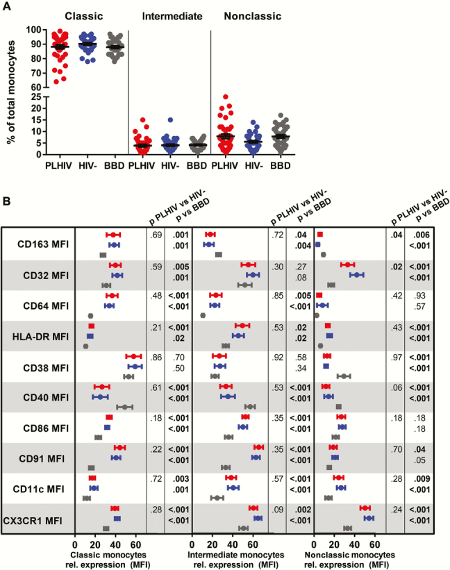
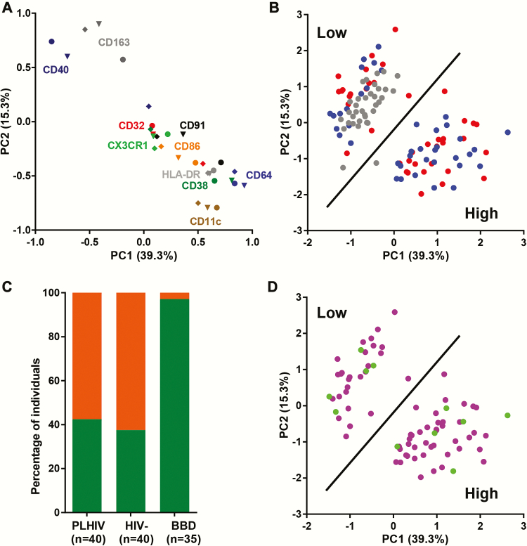
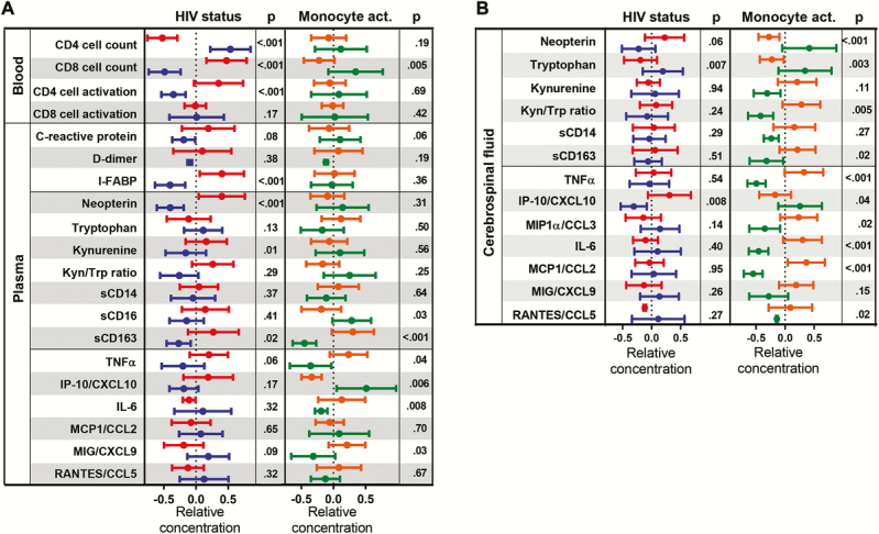
Results: People living with HIV, HIV-negative individuals, and BBD had comparable percentages of classical, intermediate, and nonclassical monocytes. Expression of CD163, CD32, CD64, HLA-DR, CD38, CD40, CD86, CD91, CD11c, and CX3CR1 on monocytes did not differ between PLHIV and HIV-negative individuals, but it differed significantly from BBD. Principal component analysis revealed that 57.5% of PLHIV and 62.5% of HIV-negative individuals had a high monocyte activation profile compared with 2.9% of BBD. Cellular monocyte activation in the COBRA cohort was strongly associated with soluble markers of monocyte activation and inflammation in the CSF.

Conclusions: People living with HIV and HIV-negative COBRA participants had high levels of cellular monocyte activation compared with age-matched BBD. High monocyte activation was predictive for inflammation in the CSF.

Keywords: CSF; HIV; immune activation; inflammation; monocyte..


Figures

Figure 1.
Figure 1.
Phenotyping of monocytes from people living with human immunodeficiency virus (PLHIV), HIV-negative Comorbidity in Relation to AIDS (COBRA) participants and blood bank donors (BBD). (a) Percentages of classical (CD14+CD16), intermediate (CD14+CD16+), and nonclassical (CD14+CD16++) monocytes of the total number of monocytes. (b) Relative expression of markers of activation (CD163, CD32, CD64, HLA-DR, and CD38), T-cell costimulation (CD40 and CD86), and adhesion (CD91, CD11c, and CX3CR1) molecules on classical, intermediate, and nonclassical monocytes. Mean fluorescence intensities (MFIs) were normalized to 100% over the classical, intermediate, and nonclassical monocyte subpopulations of PLHIV (red), HIV-negatives COBRA participants (blue), and BBD (gray) and represented as the mean and 95% confidence interval. Uncorrected P values assessed by multivariable linear regression corrected for age and gender are shown.
Figure 2.
Figure 2.
Principal component analysis of cellular markers of monocyte activation, costimulation, and adhesion data from people living with human immunodeficiency virus (PLHIV), HIV-negative Comorbidity in Relation to AIDS (COBRA) participants, and blood bank donors (BBD). (a) Loadings plot showing the relationship between the first 2 principal components (PC1, PC2) and the expression of CD163, CD32, CD64, HLA-DR, CD38, CD40, CD86, CD91, CD11c, and CX3CR1 on classical (●), intermediate (▼), and nonclassical (◆) monocytes. Each symbol shows the contribution of an individual marker to PC1 and PC2: CD163, dark gray; CD32, red; CD64, blue; HLA-DR, gray; CD38, dark green; CD40, dark blue; CD86, orange; CD91, black; CD11c, taupe; CX3CR1, green. (b) PLHIV (red), HIV-negative COBRA participants (blue), and BBD (gray) plotted based on the first 2 extracted principal components. (c) Frequency of individuals with low (green) or high (orange) cellular monocyte activation in PLHIV, HIV-negative COBRA participants, and BBD. (d) Cytomegalovirus (CMV)-positive (purple) and CMV-negative (green) COBRA participants plotted based on the first 2 extracted principal components.
Figure 3.
Figure 3.
T-cell activation and soluble markers of coagulation, gut damage, monocyte activation, and inflammation in plasma and cerebrospinal fluid (CSF). (a) Systemic markers of monocyte activation (neopterin, tryptophan, kynurenine, soluble [s]CD14, sCD16, and sCD163), inflammation (C-reactive protein, TNFα, IP-10/CXCL10, IL-6, MCP1/CCL2, MIG/CXCL9, and RANTES/CCL5), intestinal damage (I-FABP), coagulation (D-dimer), and T-cell activation (%CD38+HLA-DR+ CD4 and CD8 T cells) in people living with human immunodeficiency virus (PLHIV) (red) and HIV-negative Comorbidity in Relation to AIDS (COBRA) participants (blue) and in all COBRA participants individuals with high (orange) or low (green) cellular monocyte activation. (b) Cerebrospinal fluid concentrations of monocyte activation (neopterin, tryptophan, kynurenine, sCD14, and sCD163) and inflammation (TNFα, IP-10/CXCL10, MIP1α/CCL3, IL-6, MCP1/CCL2, MIG/CXCL9, and RANTES/CCL5) markers in PLHIV (red) and HIV-negative COBRA participants (blue) and in all COBRA participants individuals with high (orange) or low (green) cellular monocyte activation. Data were normalized by Z-score normalization and represented as the mean and 95% confidence interval. Uncorrected P values assessed by multivariable linear regression corrected for age and gender are shown.

References

    1. Guaraldi G, Orlando G, Zona S et al. Premature age-related comorbidities among HIV-infected persons compared with the general population. Clin Infect Dis 2011; 53:1120–6. - PubMed
    1. Hasse B, Ledergerber B, Furrer H et al. Morbidity and aging in HIV-infected persons: the Swiss HIV cohort study. Clin Infect Dis 2011; 53:1130–9. - PubMed
    1. Schouten J, Wit FW, Stolte IG et al. Cross-sectional comparison of the prevalence of age-associated comorbidities and their risk factors between HIV-infected and uninfected individuals: the AGEhIV cohort study. Clin Infect Dis 2014; 59:1787–97. - PubMed
    1. Deeks SG. HIV infection, inflammation, immunosenescence, and aging. Annu Rev Med 2011; 62:141–55. - PMC - PubMed
    1. Brenchley JM, Price DA, Schacker TW et al. Microbial translocation is a cause of systemic immune activation in chronic HIV infection. Nat Med. 2006; 12:1365–71. - PubMed