

Green synthesis of silver nanoparticles: Advantages of the yeast Saccharomyces cerevisiae model
- PMID: 28680992
- PMCID: PMC5490325
- DOI: 10.18869/acadpub.cmm.1.3.17
Green synthesis of silver nanoparticles: Advantages of the yeast Saccharomyces cerevisiae model
Abstract
Background and purpose: Microorganism-based synthesis of nanostructures has recently been noted as a green method for the sustainable development of nanotechnology. Nowadays, there have been numerous studies on the emerging resistant pathogenic bacteria and fungal isolates, the probable inability of bacteria and fungi to develop resistance against silver nanoparticles' (SNPs) antibacterial, antifungal, antiviral and, particularly antibacterial activities. In this study, we aim to use the yeast Saccharomycescerevisiae model for synthesis of SNPs and to investigate its antifungal activity against some isolates of Candidaalbicans.
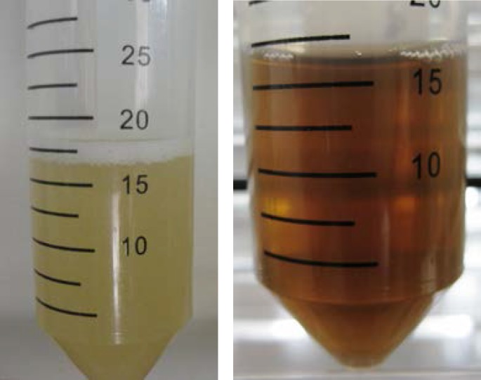
Materials and methods: A standard strain of S.cerevisiae was grown in liquid medium containing mineral salt; then, it was exposed to 2 mM AgNO3. The reduction of Ag+ ions to metal nanoparticles was virtually investigated by tracing the color of the solution, which turned into reddish-brown after 72 hours. Further characterization of synthesized SNPs was performed afterwards. In addition, antifungal activity of synthesized SNPs was evaluated against fluconazole-susceptible and fluconazole-resistant isolates of Candidaalbicans.
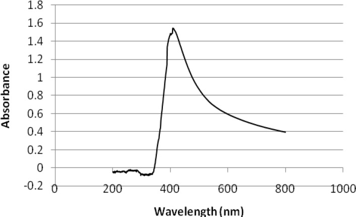
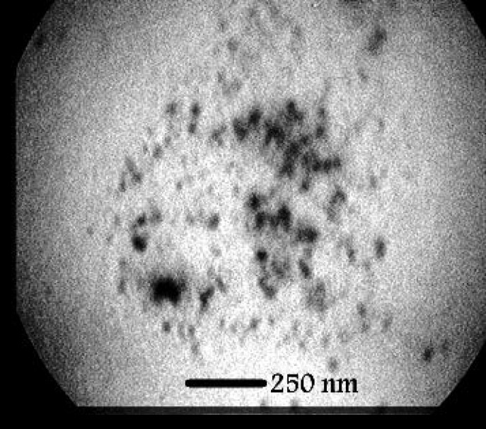
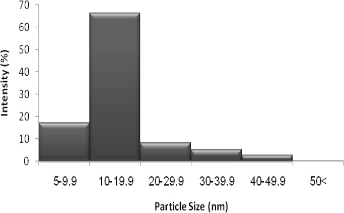
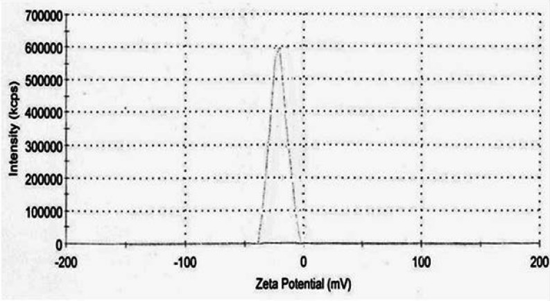
Results: The UV-vis spectra demonstrated a broad peak centering at 410 nm, which is associated with the particle sizes much less than 70 nm. The results of TEM demonstrated fairly uniform, spherical and small in size particles with almost 83.6% ranging between 5 and 20 nm. The zeta potential of SNPs was negative and equal to -25.0 (minus 25) mv suggesting that there was not much aggregation. Silver nanoparticles synthesized by S.cerevisiae, showed antifungal activity against fluconazole-susceptible and fluconazole-resistant Candida albicans isolates, and exhibited MIC90 values of 2 and 4 μg/ml, respectively.
Conclusion: The yeast S. cerevisiae model demonstrated the potential for extracellular synthesis of fairly monodisperse silver nanoparticles.
Keywords: Biosynthesis; Metal nanoparticles; Saccharomyces cerevisiae.
Figures
References
-
- Kowlgi K, Lafont U, Rappolt M, Koper G. Uniform metal nanoparticles produced at high yield in dense microemulsions. J Colloid Interf Sci. 2012;372(1):16–23. - PubMed
-
- Thakkar KN, Mhatre SS, Parikh RY. Biological synthesis of metallic nanoparticles. Nanomedicine. 2010;6(2):257–62. - PubMed
-
- Moazeni M, Shahverdi AR, Nabili M, Noorbakhsh F, Rezaie S. Green synthesis of silver nanoparticles: The reasons for and against Aspergillus parasiticus. Nanomed J. 2014;1(4):267–75.
-
- Liu L, Liu T, Tade M, Wang S, Li X, Liu S. Less is more, greener microbial synthesis of silver nanoparticles. Enzyme Microb Tech. 2014;67:53–8. - PubMed
-
- Chen X, Schluesener HJ. Nanosilver: a nanoproduct in medical application. Toxicol lett. 2008;176(1):1–12. - PubMed
LinkOut - more resources
Full Text Sources
Other Literature Sources