. 2017 Jul 6;8(7):e2915.
doi: 10.1038/cddis.2017.307.

Conservation and divergence of mitochondrial apoptosis pathway in the Pacific oyster, Crassostrea gigas

Affiliations

Conservation and divergence of mitochondrial apoptosis pathway in the Pacific oyster, Crassostrea gigas

Yingxiang Li et al. Cell Death Dis. .

Abstract

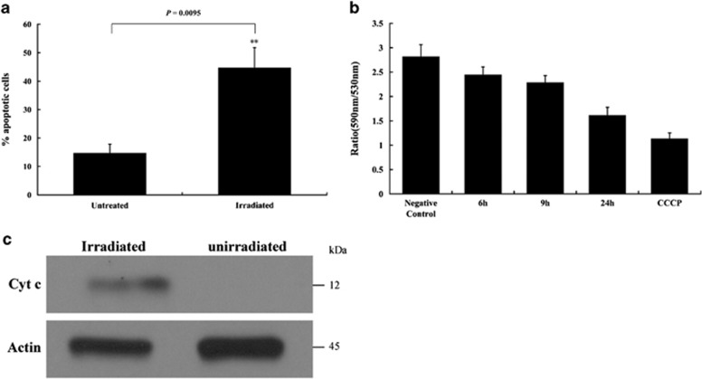
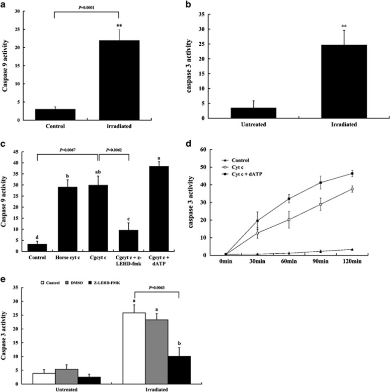
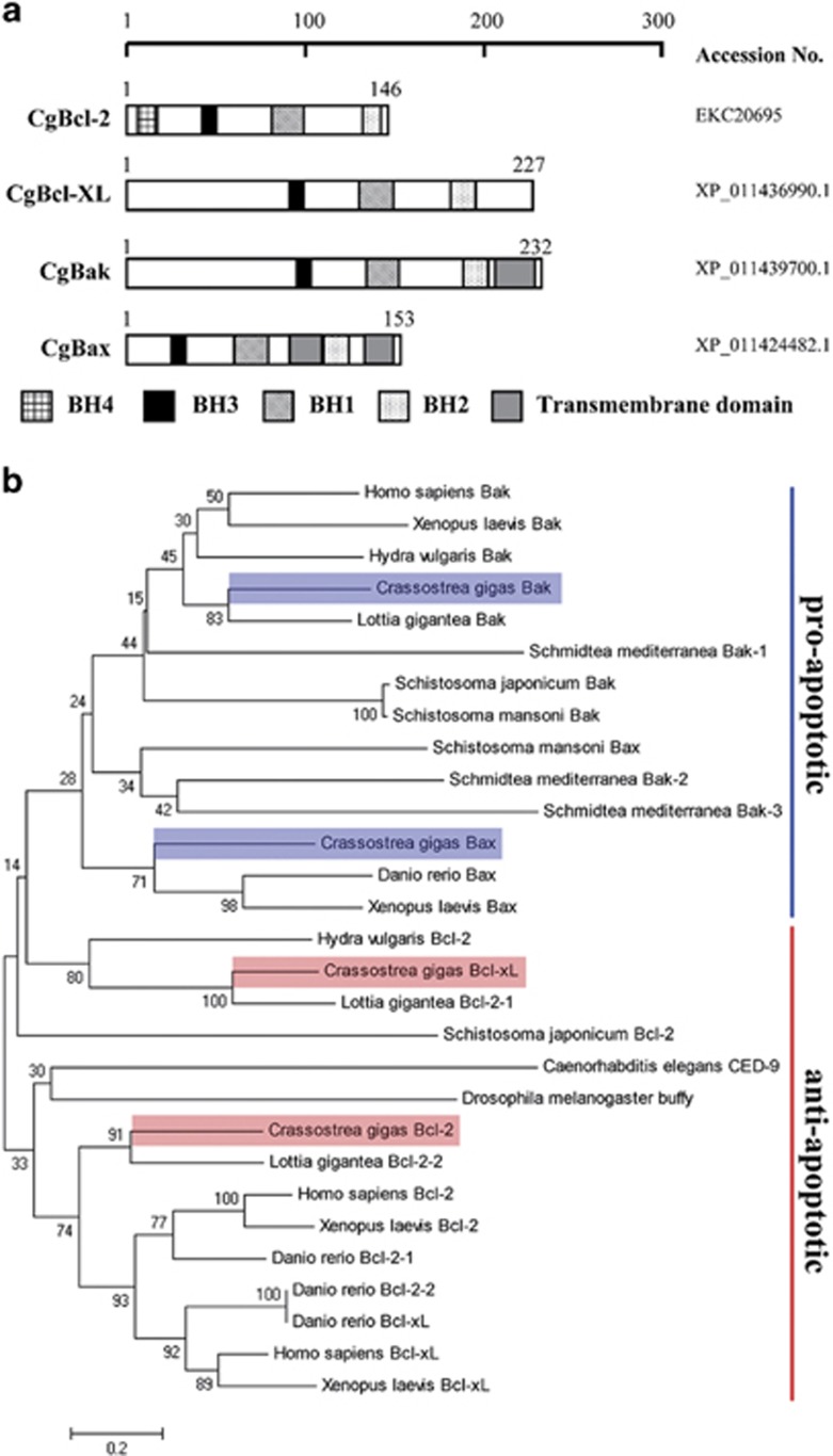
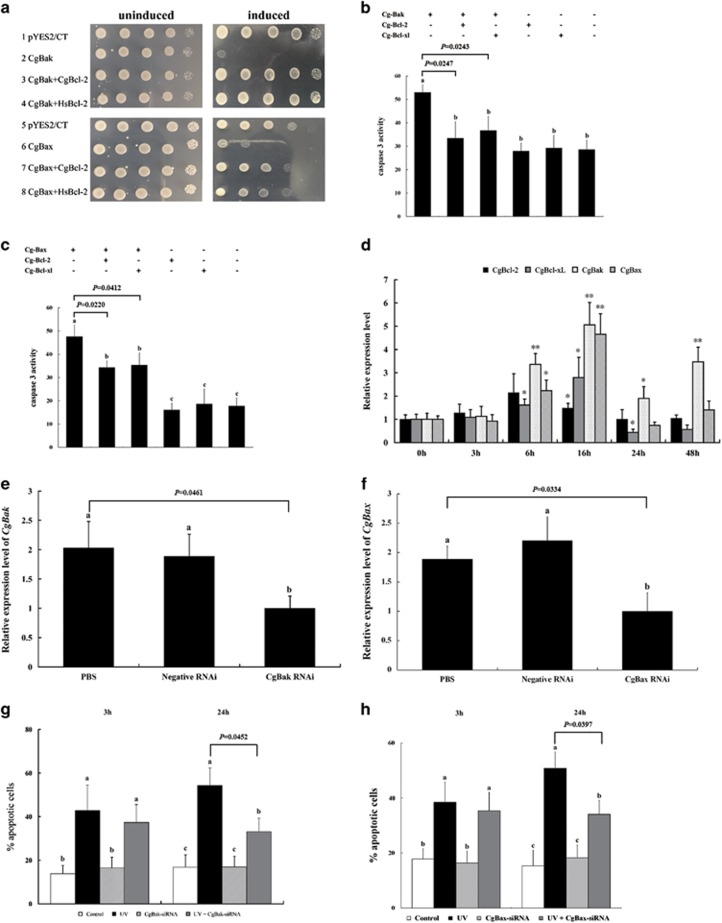
Apoptosis is considered a crucial part of the host defense system in oysters according to previous reports; however, the exact process by which this occurs remains unclear. Besides, mitochondrial apoptosis is the primary method of apoptosis in vertebrate cells, but has been poorly studied in invertebrates and is quite controversial. In this study, we investigated the molecular mechanism of mitochondrial apoptosis in the Pacific oyster Crassostrea gigas. Notably, we show that most key elements involved in the vertebrate mitochondrial apoptosis pathway - including mitochondrial outer membrane permeabilization, cytochrome c release, and caspase activation - are also present in C. gigas. In contrast, the lack of Bcl-2 homology 3-only subfamily members and apoptotic protease activating factor-1 (APAF-1) protein revealed evolutionary diversity from other phyla. Our results support that mitochondrial apoptosis in animals predates the emergence of vertebrates, but suggest that an unexpectedly diverse mitochondrial apoptosis pathway may exist in invertebrates. In addition, our work provided new clues for an improved understanding of how bivalve acclimate themselves to an inconstant environment.


Conflict of interest statement

The authors declare no conflict of interest.

Figures

Figure 1
Figure 1
Analysis of apoptosis, mitochondrial membrane potential (MMP), and cytochrome c release in UV-irradiated hemocytes of C. gigas. (a) Oyster hemocytes were irradiated with UV light and apoptosis was monitored by Annexin V/propidium iodide (PI) immunostaining at 20 h post irradiation (hpi). Data are shown as the mean±S.D. (N=6). **P<0.01. (b) Oyster hemocytes were UV-irradiated or treated with 10 μM carbonylcyanide-p-chlorophenyl hydrazone (CCCP), and hemocyte MMP was measured by JC-1 assay. Irradiated cells were sampled and measured at 6 hpi, 9 hpi, and 24 hpi. CCCP-treated and untreated/non-irradiated cells served as positive and negative controls, respectively. Lower 590/530 nm ratios refer to lower ΔΨm. N=6 per time point (including negative control and CCCP group), data are shown as the mean±S.D. (c) Cytosolic proteins were extracted from irradiated (24 hpi) and non-irradiated oyster hemocytes and analyzed by western blot using anti-cytochrome c and anti-actin antibodies
Figure 2
Figure 2
Determination of caspase 9 and caspase 3 involvement in mitochondrial apoptosis in C. gigas. (a) Caspase 9 and (b) Caspase 3 activity in oyster hemocytes was measured at 20 hpi. Non-irradiated cells were used as a negative control. Data are displayed as the mean±S.D. (N=6). **P<0.01. (c) Cytosolic extracts from oyster hemocytes were treated with cytochrome c (cyt c), Z-LEHD-FMK, and dATP. Caspase 9 activity was measured at 2 h post cyt c treatment (10 μM of horse cyt c or oyster cyt c). In Cg-cyt c+Z-LEHD-FMK group, the cytosolic extracts were preincubated with Z-LEHD-FMK (35 μM) for 1 h and then treated with Cg-cyt c. For Cg-cyt c+dATP treatment, the extracts were incubated with both Cg-cyt c and dATP (1 mM) for 2 h before activity assessment. Different letters indicate significant differences at P<0.05, whereas conditions annotated with the same letter were not significantly different. (d) Caspase 3 activity in oyster cytosolic extracts was monitored at different time points after treatment with 10 μM oyster cyt c with or without 1 mM dATP. Untreated extract served as a negative control. (e) Caspase 3 activity was examined in irradiated and non-irradiated hemocytes pretreated with Z-LEHD-FMK, DMSO vehicle, or left untreated at 20 hpi. Different small letters denote significant differences at P<0.05 while same letters denote not. All of the results in Figure 2 are shown as the mean±S.D. (N=6)
Figure 3
Figure 3
Sequence analysis of oyster Bcl-2 family proteins. (a) Small motif architecture of Bcl-2 family proteins in Crassostrea gigas. C. gigas Bcl-2 family proteins contained at least three of four conserved sequence motifs known as Bcl-2 homology domains (BH1–BH4, indicated in the figure). Cg-Bak and Cg-Bax also contained the transmembrane domain (gray box). (b) Neighbor-joining phylogenetic tree of Bcl-2 family homologs from different vertebrate and invertebrate species. The neighbor-joining tree constructed by the MEGA program was based on the sequences of four Bcl-2 family proteins in C. gigas, along with Bcl-2 family homologs from other species, including Bcl-2 homologs from Homo sapiens (NP_000624.2), Xenopus laevis (NP_001139565.1), Danio rerio (NP_001025424.1 and NP_571882.1), Drosophila melanogaster (NP_523702.1), Caenorhabditis elegans (NP_499284.1), Lottia gigantea (XP_009061217.1 and XP_009067239.1), Schistosoma japonicum (CAX69465.1), and Hydra vulgaris (NP_001274311.1); Bcl-xL homologs from Homo sapiens (CAA80661.1), Xenopus laevis (NP_001082147.1), and Danio rerio (NP_571882.1), Bak homologs from Homo sapiens (NP_001179.1), Xenopus laevis (NP_001089587.1), Lottia gigantea (XP_009064284.1), Schistosoma japonicum (CAX70134.1), Schistosoma mansoni (CCD80772.1), Schmidtea mediterranea (AEX93474.1, AEX93475.1 and AEX93476.1), and Hydra vulgaris (NP_001296708.1), and Bax homologs from Xenopus laevis (NP_001079104.1), Danio rerio (NP_571637.1), and Schistosoma mansoni (CCD81694.1)
Figure 4
Figure 4
Functional analysis of oyster Bcl-2 family proteins. (a) Plasmids expressing the indicated oyster or human Bcl-2 family proteins were transformed into Saccharomyces cerevisiae to determine their effect on yeast cell growth. Yeast were plated in 10-fold serial dilutions and induced to express the transformed genes. (b, c) HEK293T cells transfected with various plasmids (indicated in the figure) were treated with 146 μM H2O2 for 40 min. Caspase 3 activity was examined 20 hpi by spectrophotometric detection of the chromophore p-nitroaniline (pNA) after cleavage from the labeled substrate DEVD-pNA. Data are shown as the mean±S.D. (N=3). Different small letters indicate significant differences (P<0.05), whereas the same letter indicated not. (d) Expression patterns of oyster Bcl-2 family genes following irradiation were detected. Data are displayed as the mean±S.D. (N=3). *P<0.05, ** P<0.01. (e, f) Analysis of Cg-Bak and Cg-Bax mRNA levels in oyster hemocytes after siRNA transfection was used to confirm the RNAi knockdown. PBS-treated hemocytes were used as a negative control. Hemocytes in the negative RNAi group were treated with siRNA sequences as indicated in Method section. Data are displayed as the mean±S.D. (N=3). Different small letters denote significant differences (P<0.05) and the same letter denote not. (g, h) SiRNA Cg-Bak-treated and Cg-Bax-treated hemocytes were UV-irradiated and apoptosis levels were measured at 3 hpi and 24 hpi. Data are shown as the mean±S.D. (N=6). Different small letters referred to differences at P<0.05
Figure 5
Figure 5
Investigation on the apoptotic regulatory functions of Cg-Bak and Cg-Bax. (a) HeLa cells were transfected with Cg-Bak-EGFP or pEGFP-N1 empty vector and UV-irradiated or left untreated. Cell nuclei were stained with Hoechst 33342 (blue) and Alexa Fluor 633 (red) or MitoView 633 (red) to stain the cell membrane or mitochondria, respectively, as indicated in the figure. The green fluorescent signal indicated the distribution of overexpressed proteins. (b) HeLa cells were transfected with Cg-Bax-EGFP or pEGFP-N1 empty vector (green) and then UV-irradiated or left untreated. Cell nuclei were stained with Hoechst 33342 (blue). (c) Cg-Bak-EGFP and Cg-Bax-EGFP were overexpressed in HEK293T cells, which were then subjected to UV irradiation. Cytosolic and mitochondrial extracts from non-irradiated and irradiated cells were examined by western blot with anti-GFP antibody. (d) Isolated mitochondria from oyster cells were incubated with storage buffer, CaCl2 (300 μM), recombinant Cg-Bak protein (8 μM), or recombinant Cg-Bax (8 μM) at room temperature for 30 min prior to centrifugation. The distributions of cytochrome c in the supernatant (S) and precipitate (P) were then analyzed by western blot
Figure 6
Figure 6
Interactions of oyster Bcl-2 anti-apoptotic and pro-apoptotic subfamily proteins. (a) Hybridized yeast containing both tested proteins was cultured on Quadruple drop-out (QDO) plates and imaged 3–5 days later. Blue colonies indicate a potential direct interaction. (b) Names and structures of the truncated mutant proteins used in this study. The BH1 (diagonal), BH2 (dots), BH3 (solid black), BH4 (grid), and transmembrane (solid gray) domains are shown. Numbers denote the nucleic acid location. (c) Interaction between distinct truncated mutant proteins and oyster Bcl-2 family proteins detected by yeast two-hybrid system. (d) Tested proteins carrying either FLAG- or Myc tags were co-overexpressed in HEK293T cells and interactions determined by co-immunoprecipitation assays using M2 anti-FLAG antibody. (Top) anti-Myc pulldown, (Middle) western blot using anti-Myc antibody, and (Bottom) western blot using anti-FLAG antibody. (e) Interaction between distinct truncated mutant proteins and oyster Bcl-2 family proteins detected by co-IP assays
Figure 7
Figure 7
Involvement of Cg-p53 in mitochondrial apoptosis in C. gigas. (a) Expression pattern of Cg-p53 in oyster hemocytes upon UV light irradiation. (b) Effect of PFT-α treatment on Cg-p53 expression. Oyster hemocytes were incubated with 100 μM PFT-α for 1 h and Cg-p53 expression was analyzed 3 h and 6 h later. The data in (a) and (b) are shown as the mean±S.D. (N=3). (c) Apoptosis was monitored by annexin V/PI immunostaining in hemocytes following treatment as indicated. Data are displayed as the mean±S.D. (N=6). *P<0.05, **P<0.01
Figure 8
Figure 8
Role of Cg-p53 on regulating Bcl-2 family activities in mitochondrial apoptosis pathway in C. gigas. (a) Schematic diagram showing the regions of Cg-Bak, Cg-Bax, Cg-Bcl-2, and Cg-Bcl-xl considered as presumed promoter regions (gray) and subcloned into the pGL3-basic plasmid to construct the luciferase reporter. (b) Transactivation activity of Cg-p53 to Cg-Bak. The luciferase reporter containing presumed promoter region of Cg-Bak, pCMV-Cg-p53, and pRL-TK (as an internal control) were co-transfected into HEK293T cells and luciferase activity was measured 24 h later. Transactivation activity is expressed as fold increase over the control group (far left column) with no pCMV-Cg-p53. (c) Transactivation activity of Cg-p53 to Cg-Bax. (d) Transactivation activity of Cg-p53 to Cg-Bcl-2. (e) Transactivation activity of Cg-p53 to Cg-Bcl-xl. (f) Cg-Bak, and (g) Cg-Bax expression in PFT-α-treated hemocytes following irradiation. Data in (bg) are shown as the mean±S.D. (N=3). *P<0.05, **P<0.01. (h) Interactions between Cg-p53 and the four oyster Bcl-2 family proteins determined by co-IP assays in HEK293T cells. (Top) The IP samples against anti-Myc antibody. (Middle) The input samples against anti-Myc antibody. (Bottom) The input samples against anti-FLAG antibody

Similar articles

Cited by

References

    1. Guofan Z, Xiaodong F, Ximing G, Li L, Ruibang L, Fei X et al. The oyster genome reveals stress adaptation and complexity of shell formation. Nature 2012; 490: 49–54. - PubMed
    1. Francis MH, Brent F, Snimar G, Inna MS. Apoptosis as a host defense mechanism in Crassostrea virginica and its modulation by Perkinsus marinus. Fish Shellfish Immunol 2010; 29: 247–257. - PubMed
    1. Sokolova IM, Evans S, Hughes FM. Cadmium-induced apoptosis in oyster hemocytes involves disturbance of cellular energy balance but no mitochondrial permeability transition. J Exp Biol 2004; 207: 3369–3380. - PubMed
    1. Foster B, Grewal S, Graves O, Hughes FM, Sokolova IM. Copper exposure affects hemocyte apoptosis and Perkinsus marinus infection in eastern oysters Crassostrea virginica (Gmelin). Fish Shellfish Immunol 2011; 31: 341–349. - PubMed
    1. Renault T, Faury N, Barbosa-Solomieu V, Moreau K. Suppression substractive hybridisation (SSH) and real time PCR reveal differential gene expression in the Pacific cupped oyster, Crassostrea gigas, challenged with Ostreid herpesvirus 1. Dev Comp Immunol 2011; 35: 725–735. - PubMed

MeSH terms

Substances

LinkOut - more resources