. 2017 Jul 5;547(7661):E1-E3.
doi: 10.1038/nature22816.

Upholding a role for EMT in breast cancer metastasis

Affiliations

Upholding a role for EMT in breast cancer metastasis

Xin Ye et al. Nature. .
No abstract available


Conflict of interest statement

Competing Financial Interests R.A.W. is listed as an inventor on three patent applications filed by the Whitehead Institute for Biomedical Research relating to the formation of cancer stem cells through activation of an EMT program. US patent application number US 14/441,697 describes a method to use PKCα/FRA1 pathway inhibitors to target carcinoma cells that are identified by their increased expression of EMT-related genes. US patent application number US 15/307,657 describes a method to induce cancer stem cells to undergo EMT rendering them amenable to conventional treatments. US patent application number US 14/065,311 describes methods for identifying compounds and compositions that target cancer stem cells, including cancer stem cells that have undergone EMT R.A.W. is also listed as an inventor on two related patents that have been granted. US patent number US9212347 describes methods for preparing progenitor cells by inducing EMT. US patent number US9308238 describes methods and compounds for modulating and inducing EMT. R.A.W. is also a cofounder of Verastem Inc.

Figures

Extended Data Figure 1 |
Extended Data Figure 1 |. Individual channels of the stained images shown in Fig. 1b, c.
a-d, DAPI nuclear staining is shown in blue. Anti-cytokeratin staining is shown in grey. Snail-expressing cells are marked by anti-YFP staining in green (a, b). Anti-Zeb1 staining is shown in green (c, d). Anti-Fspl (a, c) and anti-Vim (b, d) staining are shown in red. Arrows indicate cytokeratin-positive tumour cells that express either the Snail (a, b) or the Zeb1 (c, d) EMT transcription factor but, at the same time, lack evident Fsp1 (a, c) or Vim (b, d) expression. Scale bars, 20 μm.
Figure 1 |
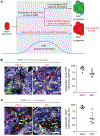
Figure 1 |. Fspl and vimentin are not universal markers of EMT programs in the MMTV-PyMT breast cancer model.
a, Schematic illustration showing the two criteria for a lineage tracing system to faithfully report EMT. b, c, Tumour sections from Snai1YFF/+;MMTV-PyMT animals were stained with DAPI as well as for cytokeratin (an epithelial marker) and either Fsp1 or vimentin together with YFP (b), which marks Snail-expressing cells, or the EMT transcription factor Zeb1 (c). Error bars represent mean ± s.d. Expression of Snail (a, green) and Zeb1 (b, green) is seen in cells that exhibit no evidence of Fsp1 or vimentin expression (red) as indicated by the arrows. Quantifications are shown in the right panels. A minimum of three different microscopic fields were counted per tumour for each marker combination. Scale bars, 20 μm.

References

    1. Fischer KR et al. Epithelial-to-mesenchymal transition is not required for lung metastasis but contributes to chemoresistance. Nature 527, 472–476 (2015). - PMC - PubMed
    1. Tan T Z. et al. Epithelial-mesenchymal transition spectrum quantification and its efficacy in deciphering survival and drug responses of cancer patients. EMBO Mol. Med 6, 1279–1293 (2014). - PMC - PubMed
    1. Ye X et al. Distinct EMT programs control normal mammary stem cells and tumour-initiating cells. Nature 525, 256–260 (2015). - PMC - PubMed
    1. Nieto MA, Huang RY, Jackson RA & Thiery JP EMT: 2016. Cell 166, 21–45 (2016). - PubMed
    1. Cano A et al. The transcription factor snail controls epithelial-mesenchymal transitions by repressing E-cadherin expression. Nat. Cell Biol. 2, 76–83 (2000). - PubMed