. 2017 Jul 6;15(7):e2001246.
doi: 10.1371/journal.pbio.2001246. eCollection 2017 Jul.

Early postnatal exposure to isoflurane causes cognitive deficits and disrupts development of newborn hippocampal neurons via activation of the mTOR pathway

Affiliations

Early postnatal exposure to isoflurane causes cognitive deficits and disrupts development of newborn hippocampal neurons via activation of the mTOR pathway

Eunchai Kang et al. PLoS Biol. .

Erratum in

Abstract

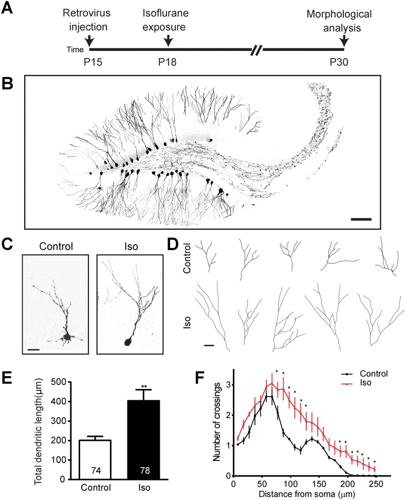
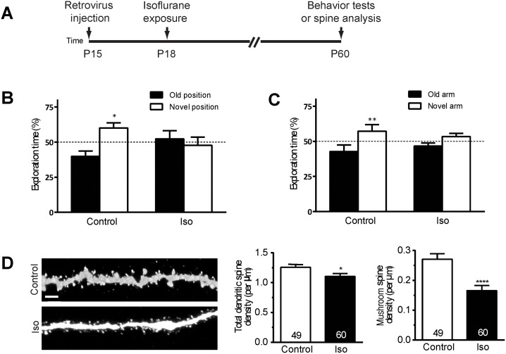
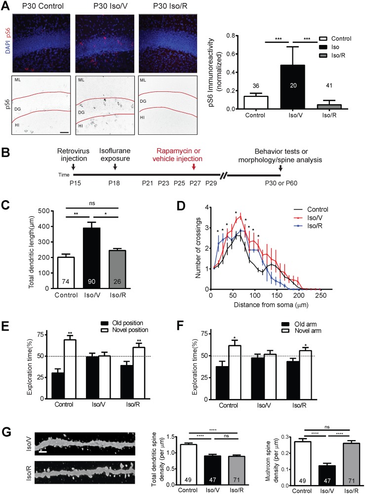
Clinical and preclinical studies indicate that early postnatal exposure to anesthetics can lead to lasting deficits in learning and other cognitive processes. The mechanism underlying this phenomenon has not been clarified and there is no treatment currently available. Recent evidence suggests that anesthetics might cause persistent deficits in cognitive function by disrupting key events in brain development. The hippocampus, a brain region that is critical for learning and memory, contains a large number of neurons that develop in the early postnatal period, which are thus vulnerable to perturbation by anesthetic exposure. Using an in vivo mouse model we demonstrate abnormal development of dendrite arbors and dendritic spines in newly generated dentate gyrus granule cell neurons of the hippocampus after a clinically relevant isoflurane anesthesia exposure conducted at an early postnatal age. Furthermore, we find that isoflurane causes a sustained increase in activity in the mechanistic target of rapamycin pathway, and that inhibition of this pathway with rapamycin not only reverses the observed changes in neuronal development, but also substantially improves performance on behavioral tasks of spatial learning and memory that are impaired by isoflurane exposure. We conclude that isoflurane disrupts the development of hippocampal neurons generated in the early postnatal period by activating a well-defined neurodevelopmental disease pathway and that this phenotype can be reversed by pharmacologic inhibition.


Conflict of interest statement

The authors have declared that no competing interests exist.

Figures

Fig 1
Fig 1. Isoflurane exposure results in overgrowth of dendritic arbors.
(A) A schematic diagram of isoflurane exposure procedure for morphology examination. (B) Sample confocal image of dentate gyrus granule cell (DGCs) infected with retrovirus expressing green florescent protein (GFP) (scale bar: 100 μm). Representative confocal images (C) and tracings (D) of individual control and isoflurane-exposed GFP+ neurons at postnatal day (P) 30 exhibiting overgrowth in the isoflurane group relative to control conditions (scale bar: 10 μm for both C and D). Summaries of total dendritic length (E) and Sholl analysis of dendritic complexity (F) of GFP+ neurons show marked overgrowth of dendritic arbors. Numbers associated with bar graph indicate the number of neurons examined from at least 5 animals per group. The same groups of neurons were examined in (E) and (F). Values represent mean ± SEM (**p < 0.01; Student t test for E and *p < 0.0001 ANOVA for F). Underlying data in S1 Data under Fig 1F tab.
Fig 2
Fig 2. Isoflurane exposure impairs spatial learning and causes a loss of dendritic spines in dentate gyrus neurons.
(A) A schematic diagram of isoflurane exposure procedure for behavior tests and spine analysis. Shown in (B and C) are summaries of the object-place recognition test (B) and the Y-maze test (C) (Control n = 12, Isoflurane n = 11; **p < 0.01, Student t test). (D) Representative processed confocal images of dendritic spines of control and isoflurane-exposed green florescent protein positive (GFP) neurons at postnatal day (P) 60 (scale bar: 2 μm). Shown on right are summary plots of total and mushroom class dendritic spine density, revealing a striking loss of mature spines. Numbers associated with the bar graph indicate the number of dendritic segments examined from at least 5 mice from each group, a total of 2,586 spines in the control group and 2,818 spines in the isoflurane group were analyzed (*p < 0.05; ****p < 0.0001, Student t test). Underlying data in S1 Data under Fig 2B-D tab.
Fig 3
Fig 3. Isoflurane exposure leads to aberrant activation of the mechanistic target of rapamycin (mTOR) signaling pathway, and pharmacological inhibition of the mTOR activities rescues deficits in behavioral tests and loss of spines.
(A) Representative confocal images of phospho-S6 (pS6) immunofluorescence at postnatal day (P) 30 in the dentate gyrus showing an increase in labeling in the isoflurane plus vehicle (Iso/V) group relative to controls and a return to baseline in the group exposed to isoflurane and subsequently treated with rapamycin, designated Iso/R. The upper panels are original confocal images with DAPI in blue and pS6 labeling in red, and the lower panels are processed for quantification with black pS6 signal on white background (ML, molecular layer; DG, dentate gyrus; HI, hilus, scale bar: 50 μm). Also shown in (A) quantification of normalized pS6 expression in the dentate gyrus granule cell layer (***p < 0.001, ANOVA, numbers in each bar represent n for images analyzed). (B) Schematic diagram of rapamycin treatment for behavior tests and spine analysis. Summaries of total dendritic length (C) and Sholl analysis of dendritic complexity (D) of GFP+ neurons show a rescue of normal dendritic arbor length and complexity with Iso/R. Values represent mean ± SEM (*p < 0.05, **p < 0.01; ANOVA for C; *p < 0.0001 ANOVA for D). Numbers in each bar represent number of cells analyzed per group, minimum of 5 animals per group). Summaries of object-place recognition test (E) and Y-maze test (F) for Iso/V and Iso/R show a recovery to near control performance with Iso/R. (Control n = 10, Iso/V n = 11, Iso/R n = 11; *: p < 0.05; **: p < 0.01, Student t test). (G) Representative confocal images of dendritic spines at P60. Scale bar: 2 μm. Shown on right are summary plots of total and mature dendritic spine density. Numbers associated with bar graph indicate the number of dendritic segments examined, a total of 2,586 spines in the control group, 1,831 spines in the isoflurane plus vehicle group, and 2,999 spines in the isoflurane plus rapamycin group were analyzed (****p < 0.0001; ns: non-significant; ANOVA, numbers in each bar represent n of dendritic segments analyzed per group, minimum of 5 animals per group). Underlying data in S1 Data under Fig 3A-G.

References

    1. DiMaggio C, Sun LS, Kakavouli A, Byrne MW, Li G. A retrospective cohort study of the association of anesthesia and hernia repair surgery with behavioral and developmental disorders in young children. J Neurosurg Anesthesiol. 2009; 21(4): 286–291. doi: 10.1097/ANA.0b013e3181a71f11 - DOI - PMC - PubMed
    1. Ing C, Dimaggio C, Whitehouse A, Hegarty MK, Brady J, von Ungern-Sternberg B, et al. Long-term Differences in Language and Cognitive Function After Childhood Exposure to Anesthesia. Pediatrics. 2012. August; peds.2011-3822; doi: 10.1542/peds.2011-3822 - DOI - PubMed
    1. Wilder RT, Flick RP, Sprung J, Katusic SK, Barbaresi WJ, Mickelson C, et al. Early exposure to anesthesia and learning disabilities in a population-based birth cohort. Anesthesiology. 2009; 110(4): 796–804. doi: 10.1097/01.anes.0000344728.34332.5d - DOI - PMC - PubMed
    1. Erasso DM, Chaparro RE, Quiroga Del Rio CE, Karlnoski R, Camporesi EM, Saporta S. Quantitative assessment of new cell proliferation in the dentate gyrus and learning after isoflurane or propofol anesthesia in young and aged rats. Brain Res. 2012; 1441: 38–46. doi: 10.1016/j.brainres.2011.11.025 - DOI - PubMed
    1. Huang L, Cichon J, Ninan I, Yang G. Post-anesthesia AMPA receptor potentiation prevents anesthesia-induced learning and synaptic deficits. Sci Transl Med. 2016; 8 (344): 344ra385. - PMC - PubMed

LinkOut - more resources