Review
. 2017 Jul 5;95(1):19-32.
doi: 10.1016/j.neuron.2017.05.021.

Toward a Neurocentric View of Learning

Affiliations
Review

Toward a Neurocentric View of Learning

Heather K Titley et al. Neuron. .

Abstract

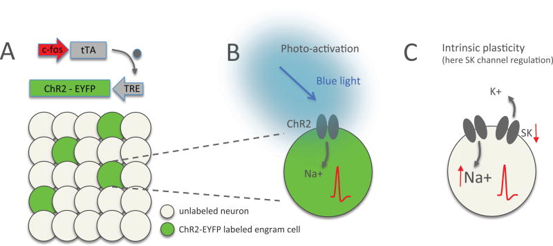
Synaptic plasticity (e.g., long-term potentiation [LTP]) is considered the cellular correlate of learning. Recent optogenetic studies on memory engram formation assign a critical role in learning to suprathreshold activation of neurons and their integration into active engrams ("engram cells"). Here we review evidence that ensemble integration may result from LTP but also from cell-autonomous changes in membrane excitability. We propose that synaptic plasticity determines synaptic connectivity maps, whereas intrinsic plasticity-possibly separated in time-amplifies neuronal responsiveness and acutely drives engram integration. Our proposal marks a move away from an exclusively synaptocentric toward a non-exclusive, neurocentric view of learning.

Keywords: Purkinje cell; cerebellum; ensemble; hippocampus; intrinsic; memory engram; neocortex; plasticity; pyramidal cell; synaptic.


Figures

Figure 1
Figure 1
Neural engram labeling / re-activation and plasticity. (A) Engram cells can be labeled by expression of ChR2-EYFP after activation of an activity-dependent promoter such as c-fos or arc. Labeled engram cells (green) can be re-activated by photo-stimulation of channelrhodopsin (ChR2). TRE: tetracycline-responsive element; tTA: tetracycline transactivator (grey dot). (B) Photo-activation of a ChR2-expressing neuron with blue light causes influx of Na+ ions and depolarization, which brings the membrane potential closer to the spike threshold and enhances the probability of spike firing. (C) Intrinsic plasticity results from a cell-autonomous modulation that similarly changes the probability of action potential generation. In the example shown here, intrinsic plasticity-related downregulation of calcium-activated, small conductance SK-type K+ channels will reduce the afterhyperpolarization following depolarizing events. As a consequence EPSPs will be enhanced and prolonged and the spike threshold will be reached more easily (EPSP amplification, faster re-depolarization toward threshold and/or reduction of the threshold itself).
Figure 2
Figure 2
Intrinsic plasticity in the primary somatosensory cortex in vivo. (A) Recording configuration. Whole-cell patch-clamp recordings are performed from L2/3 pyramidal neurons in the barrel cortex of anesthetized rats. (B) Neurons are labeled with neurobiotin (0.2%) for histological identification subsequent to the recordings. The picture shows a neurobiotin-labeled L2/3 pyramidal neuron. Scale bar: 50µm. (C)+(D) Example recordings (H.T. and C.H.; unpublished data) illustrating the bidirectionality of intrinsic excitability changes. Intrinsic plasticity is triggered by repeated injection of depolarizing current pulses (5Hz for 8s). (C) In this example recording, tetanization resulted in an increase in excitability as measured by the number of spikes evoked by test pulses. (D) Example of a neuron, in which the same tetanization protocol resulted in a depression of the spike count. Scale bars: 200ms / 20mV. Arrows indicate the time of tetanization.
Figure 3
Figure 3
Plasticity of pauses in spike firing is mediated by an intrinsic mechanism. (A) Typical trace from an in vitro Purkinje cell recording showing simple spike firing evoked by depolarizing current injection and a complex spike triggered by climbing fiber stimulation. The complex spike pause is measured as the interval between the complex spike onset and the next subsequent simple spike (gray area). (B) Top: typical traces (green) show that 5 Hz injection of depolarizing currents causes a shortening of the spike pause. In contrast, pause duration remains stable in control recordings (black traces). Bottom: time graph showing that the intrinsic plasticity protocol shortens the pause duration, while the pause remains unaltered in non-tetanized cells. (C) In SK2−/− Purkinje cells, no pause plasticity is observed. (D) Golgi staining of Purkinje cells in WT (top) and SK2−/− mice (bottom). Scale bar: 50µm. This figure is reproduced from Grasselli et al. (2016). Copyright 2016 by Elsevier.
Figure 4
Figure 4
Theoretical models of intrinsic and synaptic plasticity. (A) Left: Sketch of a simplified single neuron model showing synaptic weight adjustment by synaptic plasticity. Synaptic input weights are summed linearly and passed through a static non-linearity. Loci of plasticity are indicated in red. Right: Illustration of the hypothesis that the adjustment of synaptic weights (synaptic input map) and the plasticity of the intrinsic amplification factor can be separated in time. The example shown here depicts a face-recognition cell encoding the face of the actress Jennifer Aniston. In such highly specialized ‘concept cells’ synaptic input weights are optimally adjusted so that the neuron encodes the object, or a generalized concept of it. Intrinsic plasticity – occurring at time t+x – may enhance engram representation without altering synaptic weight ratios, but based on pre-existing connectivity. Image copyright: the authors (Editorial use license purchased from Shutterstock, Inc.). (B) – (C) Unsupervised learning. (B) Classic unsupervised synaptic plasticity rules allow a neuron to pick the direction of maximal variance in its inputs. Red dots show inputs drawn from a correlated 2D Gaussian distribution, in the space of two inputs to a neuron. The dotted line shows the initial synaptic weight vector. The total synaptic input to a neuron is given by the dot product between the input vector and the synaptic weight vector. Classic Hebbian synaptic plasticity rules – such as the Oja rule – adjust the weight vector until it picks the direction of maximal variance in the inputs, therefore performing principal component analysis (PCA). (C) The resulting distribution of total synaptic inputs is shown in red. The dotted line shows an initial static transfer function (f-I curve) that maximizes mutual information between neuron output and input. It is proportional to the cumulative distribution function of the inputs (Laughlin, 1981). Intrinsic plasticity could adjust this non-linearity until the transfer function matches the optimal one. (D) – (E) Supervised learning. (D) A classic supervised learning problem: the neuron should separate inputs into two classes (red: neuron should be active; blue: neuron should be inactive). The neuron learns to classify inputs by changing its synapses (modifying the hyperplane that separates active and inactive regions). Intrinsic plasticity can help by adjusting the neuronal threshold that measures the distance of the hyperplane from the origin. Learning can be achieved using the classic perceptron algorithm. (E) In some cases, a standard perceptron algorithm fails. In the example shown here, the neuron should learn a particular sequence of input-output associations (shown by colored dots connected by brown line). The neuron should be active in response to red inputs, but inactive for blue inputs. This example cannot be learned by a standard perceptron, because no straight line separating the blue and red dots exists. However, a bistable neuron can learn this sequence: in the bistable region (hatched region between the two thick black lines) the state of the neuron depends on the initial condition. It is active when it starts from an active state (gray shaded region), and inactive when it starts from an inactive state (white region). Intrinsic plasticity could in principle allow a neuron to become bistable and therefore allow it to solve problems that are not learnable by standard perceptrons (Clopath et al., 2013).

Similar articles

Cited by

References

    1. Acker CD, Hoyos E, Loew LM. EPSPs measured in proximal dendritic spines of cortical pyramidal neurons. eNeuro. 2016;3:e0050-15.2016. - PMC - PubMed
    1. Aizenman CD, Linden DJ. Rapid, synaptically driven increases in the intrinsic excitability of cerebellar deep nuclear neurons. Nat. Neurosci. 2000;3:109–111. - PubMed
    1. Albus JS. A theory of cerebellar function. Math. Biosciences. 1971;10:25–61.
    1. Aou S, Woody CD, Birt D. Increases in excitability of neurons of the motor cortex of cats after rapid acquisition of eye blink conditioning. J. Neurosci. 1992;12:560–569. - PMC - PubMed
    1. Armano S, Rossi P, Taglietti V, D’Angelo E. Long-term potentiation of intrinsic excitability at the mossy fiber-granule cell synapse of rat cerebellum. J. Neurosci. 2000;20:5208–5216. - PMC - PubMed

LinkOut - more resources