Review
. 2017 Jul 6;18(1):293.
doi: 10.1186/s12891-017-1654-6.

Multifocal pigmented villonodular synovitis coexisting in both the knee joint and the patella: a case report and literature review

Affiliations
Review

Multifocal pigmented villonodular synovitis coexisting in both the knee joint and the patella: a case report and literature review

Mingxuan Gao et al. BMC Musculoskelet Disord. .

Abstract

Backgroud: Pigmented villonodular synovitis (PVNS) is an uncommon entity of proliferative lesion of the synovium, presenting with different clinical signs and symptoms. PVNS rarely forms an osteolytic lesion in a bone. Here we report a unique case of PVNS with a nodular lesion in the left patella.

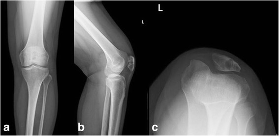
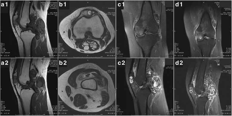
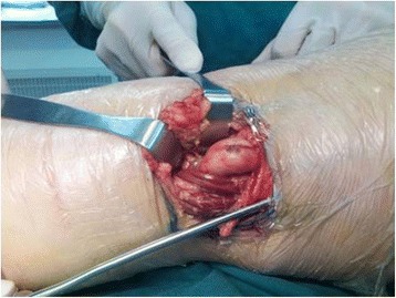
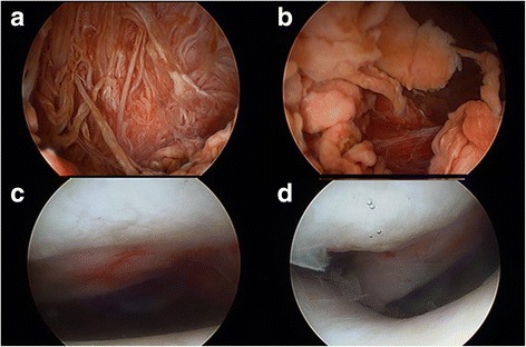
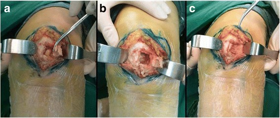
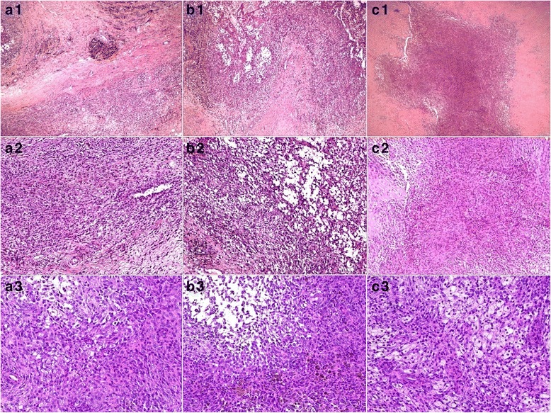
Case presentation: A 37-year-old female was referred to our hospital with complaints of ongoing left knee pain and a painful and palpable mass in her left popliteal fossa. MRI demonstrated a nodular lesion in the left patella, diffuse affected synovial tissue in the left knee and an extra-articular mass in the left popliteal fossa. After a primary diagnosis of PVNS had been established, combined arthroscopic synovectomy and open resection were performed. The postoperative pathological diagnoses of the resected mass from the popliteal fossa, the affected synovial tissue and the lesion in the patella were consistent with PVNS. At 1-year follow-up, no evidence of recurrence was noted.

Conclusions: Based on brief literature review of PNVS, we presented a very rare case of PVNS with a nodular lesion in the left patella, diffuse affected synovial tissue in the left knee and an extra-articular mass in the left popliteal fossa.

Keywords: Case report; Giant cell tumor of tendon sheath; Localized form; Multifocal lesions; Patella; Pigmented villonodular synovitis; Popliteal fossa.


Conflict of interest statement

Ethics approval and consent to participate

Not applicable.

Consent for publication

Written informed consent was obtained from the patient for the publication of this case report and any accompanying images. A copy of the written consent is available for review by the Executive Editor of this journal.

Competing interests

The authors declare that they have no competing interests.

Publisher’s Note

Springer Nature remains neutral with regard to jurisdictional claims in published maps and institutional affiliations.

Figures

Fig. 1
Fig. 1
Frontal (a), lateral (b) and the axial (c) views of the patella show a well-defined lytic lesion in the left patella, which has eroded almost all the medial half of the patella. Small degenerative osteophyte formation and slight knee joint effusion is also presented
Fig. 2
Fig. 2
Magnetic resonance imaging (MRI) reveals extensive nodular synovial proliferation in the knee, a well-defined lesion confined to the patella and a lobulated mass-like lesion in the poplitea. Sagittal T1-weighted scans (a1, a2) show inhomogeneous low signal foci and depict a clear border of the foci. Axial T2-weighted scans (b1, b2) exhibits long T2 signal of joint effusion and tissue edema. Proton density-weighted imaging (c1, c2) shows lobulated, heterogeneous lesions with high intensity of PDWI signals are in patella, the suprapatellar bursa and the posterior region of knee. Focal hypointense areas represent the hemosiderin. No destructive lighment changes and cartilage damage is detected. After the administration of gadolinium-DTPA fat suppressed T1-weighted imaging (d1, d2) illustrates a distinct enhancement of the foci. The MRI appearance of is suggestive of the PVNS diagnosis
Fig. 3
Fig. 3
A short S-shaped incision was mad over the posterior knee joint. The posterior mass in poplitea was exposed, dissected and marginally resected
Fig. 4
Fig. 4
Arthroscopic images (a, b) of the left knee indicating prolific synovium with coarse villi, heavily pigmented diffuse pigmented villonodular synovitis. No communicating link was found under arthroscopic observation (c). Although the chondromalacia changes are visible on the articular surface of patella, it seems exactly intact without any eroded hole (d)
Fig. 5
Fig. 5
A cortical window of 1.5 × 1.5 cm was made over the anteromedial aspect of the patella (a). The tumor was brownish (a) and was scooped out. On the inner side of the cavity there is a sclerotic margin (b). Bone defect in the patella was filled with iliac crest bone graft (c)
Fig. 6
Fig. 6
Photomicrographs of H&E stain samples, which were taken from the resected mass in poplitea (a1–3), the proliferated synovial tissue in the knee (b1–3) and the lesion in the patella (c1–3), respectively. Low power views (a1, b1, c1; ×40) are characterized by a prominent number of epithelioid cells with fibrous stroma. Medium power (a2, b2, c2; ×100) views demonstrates numerous mononuclear cells, scattered multinucleated giant cells and occasional cytoplasmic hemosiderin. Under higher magnification (a3, b3, c3; ×200), the mononuclear cells are round or spindled and they have minimal cytoplasm and eccentric nuclei. There are some foam cells. Some cells typically contain cytoplasmic hemosiderin granules. No mitotic figures and atypia were found. The pathological features of the lesions from different locations are very similar to one another. Histologically, all findings consistently confirmed the original diagnosis of PVNS

Similar articles

References

    1. Verspoor FG, van der Geest IC, Vegt E, Veth RP, van der Graaf WT, Schreuder HW. Pigmented villonodular synovitis: current concepts about diagnosis and management. Future Oncol. 2013;9(10):1515–1531. doi: 10.2217/fon.13.124. - DOI - PubMed
    1. Botez P, Sirbu PD, Grierosu C, Mihailescu D, Savin L, Scarlat MM. Adult multifocal pigmented villonodular synovitis-clinical review. Int Orthop. 2013;37(4):729–733. doi: 10.1007/s00264-013-1789-5. - DOI - PMC - PubMed
    1. Bouguennec N, Meyer A, Graveleau N. Localized form of pigmented villonodular synovitis of the knee: the meniscal mime. Orthop Traumatol Surg Res. 2014;100(2):251–254. doi: 10.1016/j.otsr.2013.09.017. - DOI - PubMed
    1. Young G, Marshall H. Pigmented villonodular synovitis involving bone. A case report and literature review. J Am Podiatr Med Assoc. 1989;79(7):345–347. doi: 10.7547/87507315-79-7-345. - DOI - PubMed
    1. Kindblom LG, Gunterberg B. Pigmented villonodular synovitis involving bone. Case report. J Bone Joint Surg Am. 1978;60(6):830–832. doi: 10.2106/00004623-197860060-00020. - DOI - PubMed

LinkOut - more resources