

In vivo administration of urolithin A and B prevents the occurrence of cardiac dysfunction in streptozotocin-induced diabetic rats
- PMID: 28683791
- PMCID: PMC5501434
- DOI: 10.1186/s12933-017-0561-3
In vivo administration of urolithin A and B prevents the occurrence of cardiac dysfunction in streptozotocin-induced diabetic rats
Abstract
Background: Emerging evidence suggests that specific (poly)phenols may constitute new preventative strategies to counteract cell oxidative stress and myocardial tissue inflammation, which have a key role in the patho-physiology of diabetic cardiomyopathy. In a rat model of early diabetes, we evaluated whether in vivo administration of urolithin A (UA) or urolithin B (UB), the main gut microbiota phenolic metabolites of ellagitannin-rich foods, can reduce diabetes-induced microenvironmental changes in myocardial tissue, preventing cardiac functional impairment.
Methods: Adult Wistar rats with streptozotocin-induced type-1 diabetes (n = 29) were studied in comparison with 10 control animals. Diabetic rats were either untreated (n = 9) or subjected to daily i.p. injection of UA (n = 10) or UB (n = 10). After 3 weeks of hyperglycaemia, hemodynamics, cardiomyocyte contractile properties and calcium transients were measured to assess cardiac performance. The myocardial expression of the pro-inflammatory cytokine fractalkine and proteins involved in calcium dynamics (sarcoplasmic reticulum calcium ATPase, phospholamban and phosphorylated phospholamban) were evaluated by immunoblotting. Plasma, urine and tissue distribution of UA, UB and their phase II metabolites were determined.
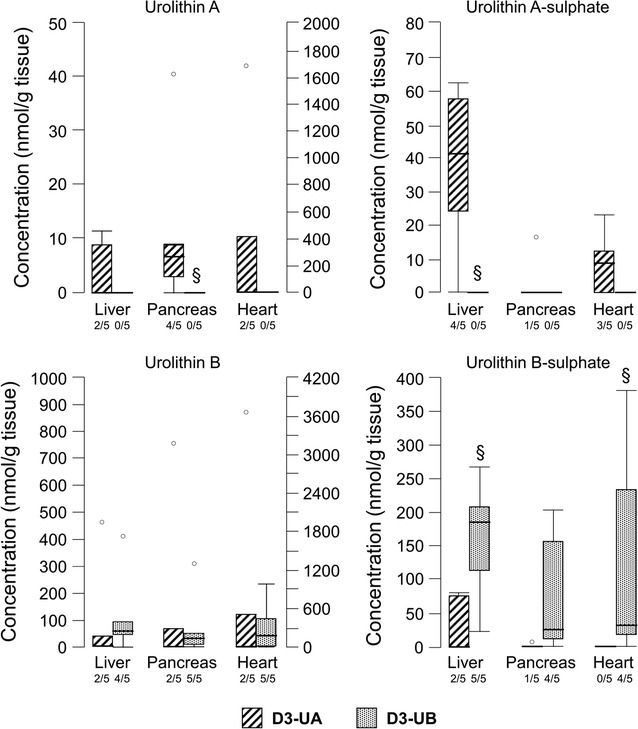
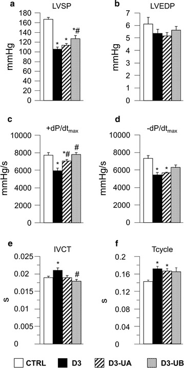
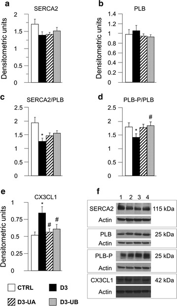
Results: In vivo urolithin treatment reduced by approximately 30% the myocardial expression of the pro-inflammatory cytokine fractalkine, preventing the early inflammatory response of cardiac cells to hyperglycaemia. The improvement in myocardial microenvironment had a functional counterpart, as documented by the increase in the maximal rate of ventricular pressure rise compared to diabetic group (+18% and +31% in UA and UB treated rats, respectively), and the parallel reduction in the isovolumic contraction time (-12%). In line with hemodynamic data, both urolithins induced a recovery of cardiomyocyte contractility and calcium dynamics, leading to a higher re-lengthening rate (+21%, on average), lower re-lengthening times (-56%), and a more efficient cytosolic calcium clearing (-32% in tau values). UB treatment also increased the velocity of shortening (+27%). Urolithin metabolites accumulated in the myocardium, with a higher concentration of UB and UB-sulphate, potentially explaining the slightly higher efficacy of UB administration.
Conclusions: In vivo urolithin administration may be able to prevent the initial inflammatory response of myocardial tissue to hyperglycaemia and the negative impact of the altered diabetic milieu on cardiac performance.
Keywords: Cardiac performance; Cardiomyocyte mechanics; Diabetes; Ellagitannins; Urolithins.
Figures

Similar articles
-
Trimethylamine-N-Oxide (TMAO)-Induced Impairment of Cardiomyocyte Function and the Protective Role of Urolithin B-Glucuronide.Molecules. 2018 Mar 1;23(3):549. doi: 10.3390/molecules23030549. Molecules. 2018. PMID: 29494535 Free PMC article.
-
Resveratrol treatment reduces cardiac progenitor cell dysfunction and prevents morpho-functional ventricular remodeling in type-1 diabetic rats.PLoS One. 2012;7(6):e39836. doi: 10.1371/journal.pone.0039836. Epub 2012 Jun 29. PLoS One. 2012. PMID: 22768138 Free PMC article.
-
Long-Term Oral Administration of Theaphenon-E Improves Cardiomyocyte Mechanics and Calcium Dynamics by Affecting Phospholamban Phosphorylation and ATP Production.Cell Physiol Biochem. 2018;47(3):1230-1243. doi: 10.1159/000490219. Epub 2018 Jun 15. Cell Physiol Biochem. 2018. PMID: 29913456
-
Hyperglycemia-induced cardiac contractile dysfunction in the diabetic heart.Heart Fail Rev. 2018 Jan;23(1):37-54. doi: 10.1007/s10741-017-9663-y. Heart Fail Rev. 2018. PMID: 29192360 Review.
-
Regulation of contractile proteins in diabetic heart.Cardiovasc Res. 1997 Apr;34(1):34-40. doi: 10.1016/s0008-6363(97)00059-x. Cardiovasc Res. 1997. PMID: 9217870 Review.
Cited by
-
Urolithin C alleviates pancreatic β-cell dysfunction in type 1 diabetes by activating Nrf2 signaling.Nutr Diabetes. 2023 Dec 1;13(1):24. doi: 10.1038/s41387-023-00253-3. Nutr Diabetes. 2023. PMID: 38040681 Free PMC article.
-
Urolithin A Attenuates Periodontitis in Mice via Dual Anti-Inflammatory and Osteoclastogenesis Inhibition: A Natural Metabolite-Based Therapeutic Strategy.Molecules. 2025 Jul 7;30(13):2881. doi: 10.3390/molecules30132881. Molecules. 2025. PMID: 40649395 Free PMC article.
-
The ellagitannin metabolite urolithin C is a glucose-dependent regulator of insulin secretion through activation of L-type calcium channels.Br J Pharmacol. 2019 Oct;176(20):4065-4078. doi: 10.1111/bph.14821. Epub 2019 Oct 10. Br J Pharmacol. 2019. PMID: 31378934 Free PMC article.
-
Blockade of Oncogenic NOTCH1 with the SERCA Inhibitor CAD204520 in T Cell Acute Lymphoblastic Leukemia.Cell Chem Biol. 2020 Jun 18;27(6):678-697.e13. doi: 10.1016/j.chembiol.2020.04.002. Epub 2020 May 7. Cell Chem Biol. 2020. PMID: 32386594 Free PMC article.
-
Trimethylamine-N-Oxide (TMAO)-Induced Impairment of Cardiomyocyte Function and the Protective Role of Urolithin B-Glucuronide.Molecules. 2018 Mar 1;23(3):549. doi: 10.3390/molecules23030549. Molecules. 2018. PMID: 29494535 Free PMC article.
References
-
- Savi M, Bocchi L, Sala R, Frati C, Lagrasta C, Madeddu D, Falco A, Pollino S, Bresciani L, Miragoli M, Zaniboni M, Quaini F, Del Rio D, Stilli D. Parenchymal and stromal cells contribute to pro-inflammatory myocardial environment at early stages of diabetes: protective role of resveratrol. Nutrients. 2016;8:729–750. doi: 10.3390/nu8110729. - DOI - PMC - PubMed
-
- Kim SJ. Herbal chrysanthemi flos, oxidative damage and protection against diabetic complications. In: Preedy Victor., editor. Diabetes: oxidative stress and dietary antioxidants. Amsterdam: Elsevier; 2014. pp. 201–211.
Publication types
MeSH terms
Substances
LinkOut - more resources
Full Text Sources
Other Literature Sources
Medical
Research Materials
