. 2017 Jul 7;10(1):23.
doi: 10.1186/s13041-017-0304-0.

Mechano growth factor, a splice variant of IGF-1, promotes neurogenesis in the aging mouse brain

Affiliations

Mechano growth factor, a splice variant of IGF-1, promotes neurogenesis in the aging mouse brain

Jason J Tang et al. Mol Brain. .

Abstract

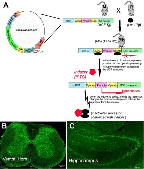
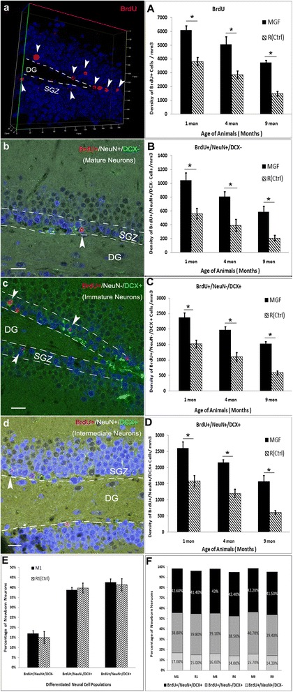
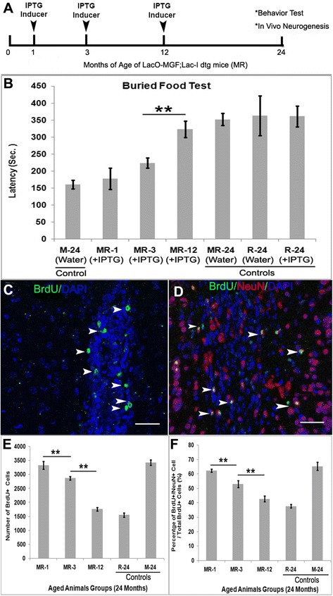
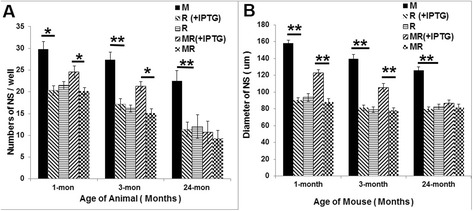
Mechano growth factor (MGF) is a splice variant of IGF-1 first described in skeletal muscle. MGF induces muscle cell proliferation in response to muscle stress and injury. In control mice we found endogenous expression of MGF in neurogenic areas of the brain and these levels declined with age. To better understand the role of MGF in the brain, we used transgenic mice that constitutively overexpressed MGF from birth. MGF overexpression significantly increased the number of BrdU+ proliferative cells in the dentate gyrus (DG) of the hippocampus and subventricular zone (SVG). Although MGF overexpression increased the overall rate of adult hippocampal neurogenesis at the proliferation stage it did not alter the distribution of neurons at post-mitotic maturation stages. We then used the lac-operon system to conditionally overexpress MGF in the mouse brain beginning at 1, 3 and 12 months with histological and behavioral observation at 24 months of age. With conditional overexpression there was an increase of BrdU+ proliferating cells and BrdU+ differentiated mature neurons in the olfactory bulbs at 24 months when overexpression was induced from 1 and 3 months of age but not when started at 12 months. This was associated with preserved olfactory function. In vitro, MGF increased the size and number of neurospheres harvested from SVZ-derived neural stem cells (NSCs). These findings indicate that MGF overexpression increases the number of neural progenitor cells and promotes neurogenesis but does not alter the distribution of adult newborn neurons at post-mitotic stages. Maintaining youthful levels of MGF may be important in reversing age-related neuronal loss and brain dysfunction.

Keywords: Aging; Insulin-like growth factor-I (IGF-I); Mechano growth factor (MGF); Neural stem cells; Neurogenesis; Neuroprotection.


Figures

Fig. 1
Fig. 1
a Scheme for generating the inducible overexpression of MGF based on the Lac operon system. Isopropyl β-D-1-thiogalactopyranoside (IPTG) is a molecular mimic of allolactose, a lactosemetabolite that triggers transcription of the Lac operon. We generated a new inducible transgene utilizing the promoter from the Huntington’s disease (HD) locus that was modified to include 2 Lac operators flanking the transcription start site to drive expression of the MGF-EGFP-Luc chimeric cDNA. When the mouse is fed with IPTG dissolved in daily water, the Lac operon is de-repressed and MGF is transcribed. The HDOS promoter drives expression specifically in neurons. GFP expression, which represents the MGF transgene, was restricted to neurons of selected area including (b) spinal cord and (c) hippocampus (negative controls not shown)
Fig. 2
Fig. 2
Identification and localization of MGF. a In the skeletal muscle, MGF-positive cells are distributed along the basal membrane of the muscle fiber. b MGF is also co-localized in MyoD-positive satellite cells along the basal membrane. Red is MGF, Green is MyoD). c MGF is expressed in the peri-nuclear region of heart muscle cells. d at the gray/white junction of the cerebral hemispheres (dotted line), MGF+ neurons were found within the cortical gray matter, rather than in white matter (WM). e in the cortex MGF-positive neurons were round with very short processes. f In the lateral ventricular (LV) region, MGF was distributed along the wall of LV. g under higher magnification, MGF-positive cells exhibited long branched cellular processes like astrocytes. h In the hippocampus MGF positive cells were distributed at the border of the of the subgranular zone (SGZ)and dentate gyrus (dotted line) which is an area of active adult neurogenesis. i A small number of rounded MGF positive neurons were also observed in the striatum. j In the hypothalamus cells were detected in the paraventricular (V3) area. k Numerous cells were also seen in ventral pallidum area of the basal ganglia. (Red is MGF, Blue is DAPI, Scale bar, (in A-K) 100 um)
Fig. 3
Fig. 3
Characterization of endogenous MGF-expressing cells in the brain. a In the SVZ, MGF positive cells also express GFAP, which is a marker of neural stem cells in the SVZ. b Immature neurons in the SVZ were positive for both DCX and (c) PSA-NCAM expression and co-expressed MGF. These double positive neurons were in the RMS. d In the hippocampus, astrocyte-like MGF positive neurons did not express Nestin in the radial-glial like neural progenitors (type-1 NPCs) in DG area. e Nestin-positive amplifying neural progenitors in DG were positive for MGF in SGZ. f MGF was also expressed in the cytoplasm of β-Tubulin III positive neurons in the neurogenic region of DG. β-Tubulin III is one of the earliest markers seen in immature neurons. g BrdU-positive proliferating cells are also co-localizing with MGF in SVZ of LV in the brain. [Red is MGF, Green is GFAP (in a), DCX (in b), PSA-NCAM (in c), Nestin (in e), β-Tubulin III (in f) and BrdU (in g)]. (Scale bar (in a-g) 100 um)
Fig. 4
Fig. 4
Regulation of MGF in the proliferation and maturation of newborn neurons in the adult hippocampus. a Representative Z stack/3D structure of the dentate gyrus (DG) of the hippocampus (40X) showing BrdU-positive proliferating cells in the subgranular zone (SGZ). (a) BrdU density between MGF transgenic mice and the age-matched control mice; MGF overexpression enhances the cell proliferation. b Representative image of new adult-born mature neurons in the SGZ labeled with BrdU+/NeuN+/DCX- . (b) MGF overexpression increases the density of new adult-born mature neurons, (c, c) new adult-born immature neurons labeled with BrdU+/NeuN−/DCX+. (d, d) differentiated intermediate neurons in transition from immature to mature neurons at 1,4 and 9-month-old,compared to all age-matched control mice. (e) At the age of 1 month about 15% of newborn neurons are mature, about 40% are immature and about 42% are overlapping between mature and immature. However, compared to the control, the MGF transgenic mice have no difference regarding the distribution of newborn neurons at different maturation stages. (f) The percentage of the newborn neurons at different maturation stages are very similar between MGF transgenic and control mice when they are aging. (Error bars indicate ± SEM. * p < 0.01, N = 4 each group); (scale bar 20um)
Fig. 5
Fig. 5
MGF overexpression preserved age-related Olfactory deficits and enhanced neural stem cell proliferation and differentiation. a Three groups of MR mice were separately fed with IPTG inducer in the daily water beginning at 1,3,12 month old (MR-1,MR-3,MR-12) and terminated at the age of 24 months old followed by function analysis and in vivo neurogenesis analysis. b Among all these 24-month-old mice, mice with constitutive overexpression of MGF throughout life (M-24) or induced overexpression of MGF from 1 month (MR-1) or 3 months (MR-3) of age had significantly improved olfactory function compared to the control mice of MR dtg mice (MR-24) with water (no IPTG) or MR dtg mice that began IPTG induction at 12 months (MR-12). MR-1, MR-3 and M-24 mice found buried food more rapidly. There was no difference among other control groups (n = 4–8 mice per group; bars represent mean ± SEM). The newborn neurons in olfactory bulb were quantitated after immunofluorescence stains. c Representative confocal images showing BrdU+ proliferating cells in the SVZ (d) differentiated mature granular neurons in olfactory bulb. e Quantification of total BrdU+ cells of olfactory bulb (f) and the percentage of newly generated granular neurons (BrdU+/NeuN+) from the total BrdU+ proliferating cells in olfactory bulb were quantitated. Early induced overexpression of MGF in the brain beginning at 1 month and 3 months had significantly enhanced the total number of BrdU+ proliferating cells and the average percentage of newly differentiated granular neurons (BrdU+/NeuN+) in the olfactory bulb by the age of 24 months. However, later overexpression of MGF induced at the age of 12 months had increased the percentage but not significantly compared to the control mice(R mice). Green(BrdU), Red(NeuN), Blue(Nuclear). (Error bars indicate ± SEM. **p < 0.01. Scale bar = 50 um
Fig. 6
Fig. 6
In vitro cell culture of SVZ-derived neurosphere (NS). Comparison of average NS numbers per well (a) and NS size at 7DIV (b). NSCs from M mice have significantly highest number and biggest size of NS at all age points (*p < 0.05,**p < 0.01), while NSC from R mice have no difference in neither sphere number nor the size (p > 0.05). IPTG induction in the MR mice-derived NSCs at age of 1 and 3 month, would significantly result in increasing of NS number and size (**p < 0.01)

Similar articles

Cited by

References

    1. Ming GL, Song H. Adult neurogenesis in the mammalian brain: significant answers and significant questions. Neuron. 2011;70:687–702. doi: 10.1016/j.neuron.2011.05.001. - DOI - PMC - PubMed
    1. Gage FH, Temple S. Neural stem cells: generating and regenerating the brain. Neuron. 2013;80:588–601. doi: 10.1016/j.neuron.2013.10.037. - DOI - PubMed
    1. Gage FH. Mammalian neural stem cells. Science. 2000;287:1433–1438. doi: 10.1126/science.287.5457.1433. - DOI - PubMed
    1. Doetsch F. A niche for adult neural stem cells. Curr Opin Genet Dev. 2003;13:543–550. doi: 10.1016/j.gde.2003.08.012. - DOI - PubMed
    1. Spalding KL, Bergmann O, Alkass K, Bernard S, Salehpour M, Huttner HB, et al. Dynamics of hippocampal neurogenesis in adult humans. Cell. 2013;153:1219–1227. doi: 10.1016/j.cell.2013.05.002. - DOI - PMC - PubMed

MeSH terms