Review
. 2017:109:223-264.
doi: 10.1016/bs.apcsb.2017.04.008. Epub 2017 Jun 5.

Sortase Transpeptidases: Structural Biology and Catalytic Mechanism

Affiliations
Review

Sortase Transpeptidases: Structural Biology and Catalytic Mechanism

Alex W Jacobitz et al. Adv Protein Chem Struct Biol. 2017.

Abstract

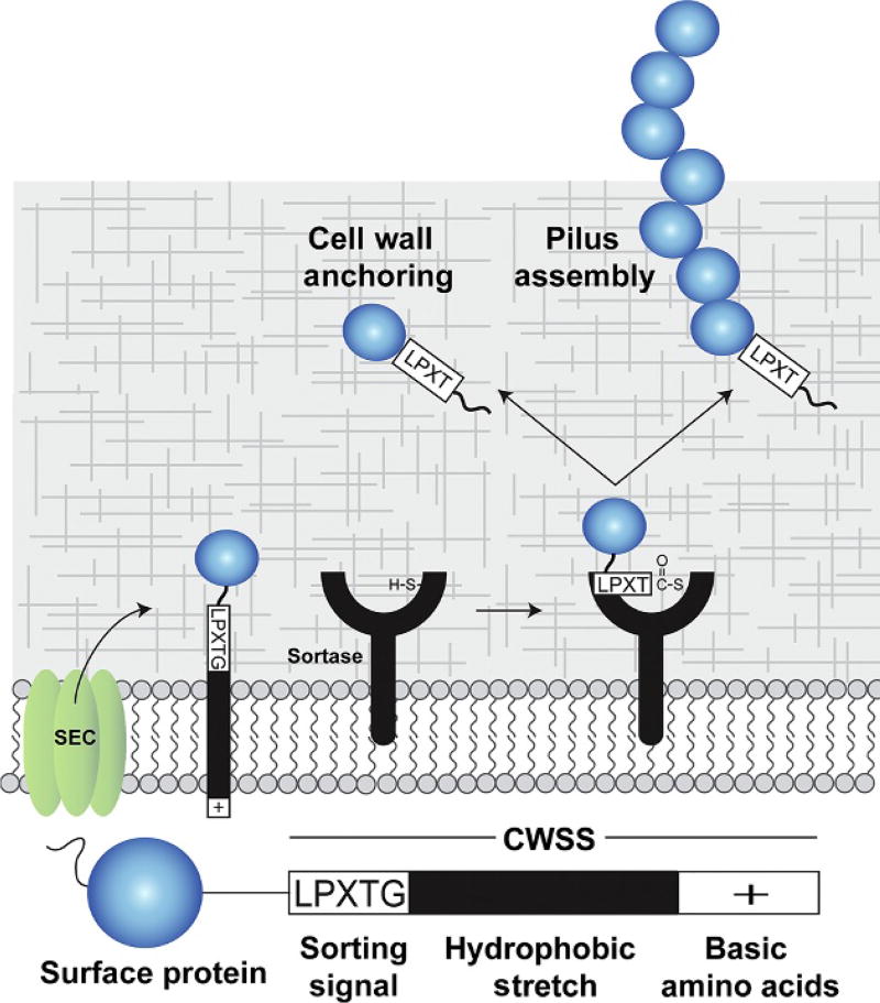
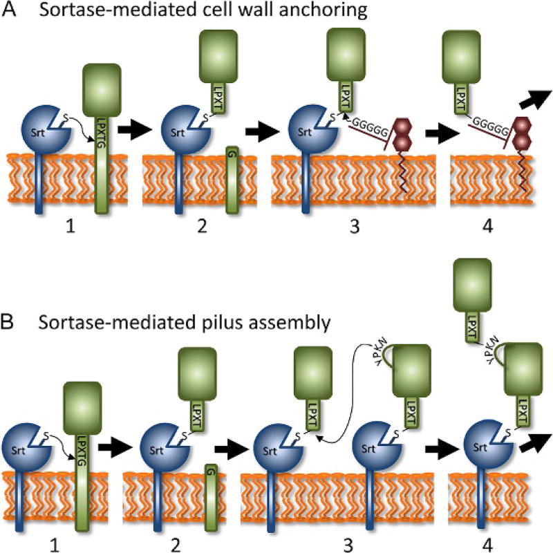
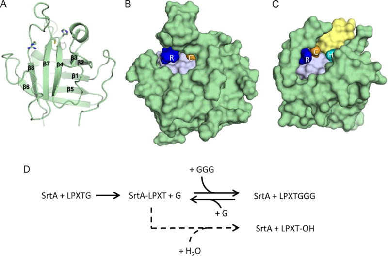
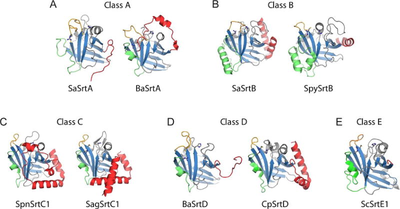
Gram-positive bacteria use sortase cysteine transpeptidase enzymes to covalently attach proteins to their cell wall and to assemble pili. In pathogenic bacteria sortases are potential drug targets, as many of the proteins that they display on the microbial surface play key roles in the infection process. Moreover, the Staphylococcus aureus Sortase A (SaSrtA) enzyme has been developed into a valuable biochemical reagent because of its ability to ligate biomolecules together in vitro via a covalent peptide bond. Here we review what is known about the structures and catalytic mechanism of sortase enzymes. Based on their primary sequences, most sortase homologs can be classified into six distinct subfamilies, called class A-F enzymes. Atomic structures reveal unique, class-specific variations that support alternate substrate specificities, while structures of sortase enzymes bound to sorting signal mimics shed light onto the molecular basis of substrate recognition. The results of computational studies are reviewed that provide insight into how key reaction intermediates are stabilized during catalysis, as well as the mechanism and dynamics of substrate recognition. Lastly, the reported in vitro activities of sortases are compared, revealing that the transpeptidation activity of SaSrtA is at least 20-fold faster than other sortases that have thus far been characterized. Together, the results of the structural, computational, and biochemical studies discussed in this review begin to reveal how sortases decorate the microbial surface with proteins and pili, and may facilitate ongoing efforts to discover therapeutically useful small molecule inhibitors.

Keywords: Mechanism; Sortase; Structure; Transpeptidase.


Figures

Fig. 1
Fig. 1
Sortase enzymes attach proteins to the cell wall and assemble pili. (A) Overview of anchoring and pilus assembly reactions. A protein that is to be displayed (blue) contains an N-terminal secretion signal and a C-terminal cell wall sorting signal (CWSS). The CWSS contains an LPXTG-like sorting signal sequence that is processed by the sortase, a nonpolar polypeptide segment (black), and a C-terminal segment of positively charged residues (+). After secretion through the Sec translocon, the protein remains embedded in the lipid bilayer via the nonpolar segment within the CWSS. The sortase enzyme then cleaves between the threonine and glycine residues to form a sortase–protein thioacyl intermediate in which the active site cysteine is covalently linked to the carbonyl carbon atom of the threonine. There are two basic types of sortases: (1) cell wall anchoring sortases that attach protein to the crossbridge peptide of the cell wall and (2) pilin polymerase sortases that covalently link pilin subunits together via lysine–isopeptide bonds. In both cases, the enzymes function as transpeptidases. Some sortases are capable of performing both functions, attaching proteins to the cell wall and polymerizing pili.
Fig. 2
Fig. 2
Mechanism of cell wall protein anchoring and pilus assembly. Sortases perform two basic functions in bacteria: (1) attach proteins to the cell wall and (2) join proteins together to construct pili. (A) In the cell wall anchoring reaction, the sortase and substrate are both membrane bound. The reaction occurs via four distinct steps. Sortase first recognizes a sorting signal motif within the CWSS and nucleophilically attacks the threonine’s carbonyl carbon atom via its active site cysteine residue (for demonstration purposes the LPXTG sorting signal recognized by class-A type enzymes is shown, step 1). The LPXTG sorting signal is then cleaved to produce a sortase–substrate thioacyl intermediate (step 2). Next, the crossbridge peptide from a lipid II molecule nucleophilically attacks the thioacyl intermediate (step 3). Lastly, a new peptide bond is formed between the lipid II molecule and surface protein to produce a protein–lipid II intermediate that is incorporated into cell wall by the transglycosylation and transpeptidation reactions that synthesize the peptidoglycan (step 4). (B) In the pilus assembly reaction, steps 1–2 produce a sortase–substrate thioacyl intermediate, similar to the cell wall anchoring reaction. In this reaction, the sortase recognizes a pilin protein that contains a CWSS. However, a lysine residue within the pilin motif from an adjacent pilin protein performs the nucleophilic attack on the thioacyl intermediate (step 3). A new protein–protein isopeptide bond is formed that covalently links the pilin proteins (step 4). This assembly process is repeated to build an isopeptide-linked pilus shaft that contains multiple pilin proteins. Depending on the type of pilus, distinct tip and base pilin proteins can be located at the ends of the pilus shaft, which are incorporated through a similar mechanism and involve covalent linkages via lysine-derived isopeptide bonds. Finally, the intact pilus is attached to the cell wall via sortase-catalyzed attachment of the pilus to lipid II, similar to cell wall protein display. Some sortases are capable of performing both functions, attaching proteins to the cell wall and functioning as pilin polymerases.
Fig. 3
Fig. 3
Structure and transpeptidation reaction of representative class A sortases. (A) S. aureus SrtA (SaSrtA) NMR structure (cartoon), showcasing an eight-stranded β-barrel with active site His120, Cys184, and Arg197 residues (sticks). (B) SaSrtA NMR structure (green surface) with active residues Arg (blue) and Cys (orange). The active site His is occluded by a closed β7/β8 loop, and there is no obvious groove for a full-length peptide to exit the active site. (C) S. pyogenes SrtA (SpySrtA) structure (green surface) with active site Arg (blue), Cys (orange), and His (cyan) residues. An open β7/β8 loop creates a clear channel that can be seen running between active Cys and His residues, indicating the potential exit channel (yellow) for the full-length peptide substrate. (D) In vitro, SaSrtA catalyzes a reversible transpeptidation reaction (top, indicated by solid arrows) in which it joins LPXTG and (Gly)3 peptides. In the absence of glycine oligopeptide, SaSrtA acts a protease and cleaves the LPXTG peptide between its threonine and glycine residues (bottom, indicated by a dashed arrow). In this spurious pathway, a water molecule, instead of lipid II, performs the second nucleophilic attack to cleave the thioacyl bond between sortase and substrate, thereby hydrolyzing the peptide. On the cell surface, hydrolysis is presumably undesirable, as proteolysis separates the protein from its membrane anchor, releasing it from the microbe. Transpeptidation occurs faster than the rate of proteolysis in vitro, making SaSrtA a valuable bioconjugation reagent (kcat = 0.28 ± 0.02 and 0.086 ± 0.015 s−1, respectively). Although all sortases are thought to catalyze transpeptidation reactions on the cell surface, this activity has only been reconstituted in vitro for a few sortases in addition to SaSrtA (listed in Table 2).
Fig. 4
Fig. 4
Structural variation by class of sortase. Sortases representative of the major themes seen for each class are displayed (cartoon) with active site residues (sticks). The hallmark sortase β-barrel (blue) and major sources of structural variability are highlighted, including N-terminus (red), β6/β7 loop (green), and β7/β8 loop (orange). Panels A–E show representative class A–E enzymes, respectively.
Fig. 5
Fig. 5
Sorting signal recognition. (A) The SaSrtA–LPAT* complex. (B) The SaSrtB–NPQT* complex. (C) The BaSrtA–LPAT* complex, shown with N-terminal appendage removed from view for clarity. Enzymes are shown as surface representations with SrtA types in light green and SaSrtB in light blue, substrate mimics are shown as gray sticks. Active site Cys and Arg residues are shown as gold and blue surfaces, respectively. (D) Conserved recognition sites for sortase enzymes. Left, SaSrtA shown as a transparent surface representation with recognition subsites determined from the combination of sortase structures color coded as follows: S4 is shown in red, S3 in orange, S2 in green, and S1 in magenta, and active site Arg in blue, Cys in gold, and His in cyan. Right, Cartoon diagram of SaSrtA with secondary structure elements that contribute to substrate binding labeled for clarity.
Fig. 6
Fig. 6
The substrate-stabilized oxyanion hole. The energy minimized model of the SaSrtB-NPQT thioacyl intermediate displayed with SaSrtB (light blue cartoon), residues in the active site and oxyanion hole (sticks), and NPQT substrate (gray sticks). The side chain hydroxyl of the substrate’s P1 Thr residue and backbone carbonyl participate in a hydrogen bonding network with the active site Arg, and the backbone amide of Glu224 that together build an oxyanion hole to stabilize the high energy tetrahedral reaction intermediates. Reproduced from Jacobitz, A. W., Wereszczynski, J., Yi, S. W., Amer, B. R., Huang, G. L., Nguyen, A. V., et al. (2014). Structural and computational studies of the Staphylococcus aureus Sortase B-substrate complex reveal a substrate-stabilized oxyanion hole, The Journal of Biological Chemistry, 289, 88918902, p. jbc.M113.509273.
Fig. 7
Fig. 7
Molecular mechanism of sortase enzymes. The active site of sortase consists of a His–Cys–Arg triad, and in its active form, the His and Cys residues form a thiolate–imidazolium ion-pair (A). The reaction begins with recognition of an appropriate sorting signal (here, the LPXTG sorting signal for SrtA types is shown), and the active site Cys residue performs nucleophilic attack on the carbonyl carbon at the substrate’s P1 position (B). An oxyanion tetrahedral intermediate is stabilized by the nearby Arg residue that is likely oriented by interactions with the side chain of the substrate’s P1 residue, which is a threonine in over 95% of all substrates (C). The active His residue concomitantly donates a proton to the leaving group, and the tetrahedral transition state then collapses to form a semistable, thioacyl intermediate between the substrate’s P1 residue and the active site Cys (D). Next, the secondary substrate (here shown as lipid II used by cell wall anchoring sortases) enters the active site, where its terminal amine is deprotonated by the active His residue before performing nucleophilic attack on the carbonyl carbon in the thioacyl bond (E); this second tetrahedral intermediate (F) collapses to form a peptide bond between the two substrates, and the product is finally released to leave the regenerated active site (A).

References

    1. Amer BR, Macdonald R, Jacobitz AW, Liauw B, Clubb RT. Rapid addition of unlabeled silent solubility tags to proteins using a new substrate-fused sortase reagent. Journal of Biomolecular NMR. 2016;64(3):197–205. - PMC - PubMed
    1. Antos JM, Truttmann MC, Ploegh HL. Recent advances in sortase-catalyzed ligation methodology. Current Opinion in Structural Biology. 2016;38:111–118. - PMC - PubMed
    1. Aulabaugh A, Ding W, Kapoor B, Tabei K, Alksne L, Dushin R, et al. Development of an HPLC assay for Staphylococcus aureus sortase: Evidence for the formation of the kinetically competent acyl enzyme intermediate. Analytical Biochemistry. 2007;360(1):14–22. - PubMed
    1. Bentley ML, Gaweska H, Kielec JM, McCafferty DG. Engineering the substrate specificity of Staphylococcus aureus Sortase A. The beta6/beta7 loop from SrtB confers NPQTN recognition to SrtA. The Journal of Biological Chemistry. 2007;282(9):6571–6581. - PubMed
    1. Bentley ML, Lamb EC, McCafferty DG. Mutagenesis studies of substrate recognition and catalysis in the Sortase A transpeptidase from Staphylococcus aureus. The Journal of Biological Chemistry. 2008;283(21):14762–14771. - PMC - PubMed

MeSH terms