Comparative Study
. 2017 Aug 11;292(32):13168-13185.
doi: 10.1074/jbc.M117.794917. Epub 2017 Jul 6.

Kinetic processivity of the two-step oxidations of progesterone and pregnenolone to androgens by human cytochrome P450 17A1

Affiliations
Comparative Study

Kinetic processivity of the two-step oxidations of progesterone and pregnenolone to androgens by human cytochrome P450 17A1

Eric Gonzalez et al. J Biol Chem. .

Abstract

Cytochrome P450 (P450, CYP) 17A1 plays a critical role in steroid metabolism, catalyzing both the 17α-hydroxylation of pregnenolone and progesterone and the subsequent 17α,20-lyase reactions to form dehydroepiandrosterone (DHEA) and androstenedione (Andro), respectively, critical for generating glucocorticoids and androgens. Human P450 17A1 reaction rates examined are enhanced by the accessory protein cytochrome b5 (b5), but the exact role of b5 in P450 17A1-catalyzed reactions is unclear as are several details of these reactions. Here, we examined in detail the processivity of the 17α-hydroxylation and lyase steps. b5 did not enhance reaction rates by decreasing the koff rates of any of the steroids. Steroid binding to P450 17A1 was more complex than a simple two-state system. Pre-steady-state experiments indicated lag phases for Andro production from progesterone and for DHEA from pregnenolone, indicating a distributive character of the enzyme. However, we observed processivity in pregnenolone/DHEA pulse-chase experiments. (S)-Orteronel was three times more inhibitory toward the conversion of 17α-hydroxypregnenolone to DHEA than toward the 17α-hydroxylation of pregnenolone. IC50 values for (S)-orteronel were identical for blocking DHEA formation from pregnenolone and for 17α-hydroxylation, suggestive of processivity. Global kinetic modeling helped assign sets of rate constants for individual or groups of reactions, indicating that human P450 17A1 is an inherently distributive enzyme but that some processivity is present, i.e. some of the 17α-OH pregnenolone formed from pregnenolone did not dissociate from P450 17A1 before conversion to DHEA. Our results also suggest multiple conformations of P450 17A1, as previously proposed on the basis of NMR spectroscopy and X-ray crystallography.

Keywords: cytochrome P450; enzyme kinetics; oxidation-reduction (redox); pre-steady-state kinetics; steady-state kinetics; steroid metabolism; steroidogenesis.


Conflict of interest statement

The authors declare that they have no conflicts of interest with the contents of this article

Figures
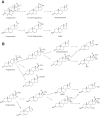
Figure 1.
Figure 1.
Oxidations catalyzed by P450 17A1. A, major classical reactions; B, expanded repertoire of reactions (14–16).
Figure 2.
Figure 2.
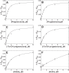
Steady-state kinetics of major reactions catalyzed by P450 17A1 and the effect of b5. The data points are means of duplicate assays (± range). Fitting was done by non-linear regression analysis of hyperbolic data in GraphPad Prism. See Table 1 for calculated values. A, pregnenolone (preg); B, 17α-OH pregnenolone; C, progesterone (prog); D, 17α-OH progesterone.
Figure 3.
Figure 3.
Binding of substrates and products to P450 17A1. Absorbance measurements were made following mixing in a stopped-flow spectrophotometer as described in detail under “Experimental procedures.” At least three data points were collected at each ligand concentration, and the points were fit to hyperbolic curves using Prism software and non-linear regression analysis (S.E. calculated from curve fitting). A, pregnenolone (Kd 0.37 ± 0.03 μm); B, progesterone (Kd 0.47 ± 0.04 μm); C, 17α-OH pregnenolone (Kd 0.52 ± 0.10 μm); D, 17α-OH progesterone (Kd 0.95 ± 0.08 μm); E, DHEA (Kd 1.7 ± 0.2 μm); F, Andro (Kd 19 ± 9 μm).
Figure 4.
Figure 4.
(S)-Orteronel binding to P450 17A1. P450 17A1 (2 μm) and (S)-orteronel (10 μm) in 100 mm potassium phosphate buffer (pH 7.4) were mixed together in the stopped-flow spectrophotometer. A, series of spectra collected at 1-ms intervals over a total period of 1.0 s (only a subset is shown). B, calculated beginning and ending spectra after SVD analysis using a two-species model in the OLIS GlobalWorks® software. C, SVD analysis of data for disappearance of starting species and appearance of final species, along with OLIS EV (Eigenvector) absorbance and OLIS normalized experimental data. D, eigenvector (solid line) and normalized experimental data. E, residuals analysis of fit to a rate of 6.1 s−1 (from C and D).
Figure 5.
Figure 5.
Estimation of koff rate for 17α-OH pregnenolone from P450 17A1 using (S)-orteronel trapping. The experiment of Fig. 4C was repeated except that the P450 17A1 in the starting system was bound in a 1:1 molar complex with 17α-OH pregnenolone, and the scan was extended to 10 s using the averaging mode (62 scans/s). A, series of spectra collected at 1-ms intervals over a total period of 1.0 s (only a subset is shown). B, calculated beginning and ending spectra after SVD analysis using a two-species model in the OLIS GlobalWorks® software. C, SVD analysis of data for disappearance of starting species and appearance of final species, along with OLIS EV (Eigenvector) absorbance and OLIS normalized experimental data. Under these conditions (4 μm P450 17A1 (E) and 4 μm 17α-OH pregnenolone (S) and using the Kd value of 0.52 μm (Fig. 3), 70% of the P450 17A1 is in the substrate-bound form (Kd = [E]free[S]free/[ES]). The calculated koff rate was 0.4 s−1 (D, with the residuals shown in E). This is a single experiment; for the entire set of averaged experiments see Table 2.
Figure 6.
Figure 6.
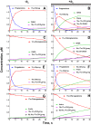
“Single-turnover” kinetics of 1:1 molar complexes of P450 17A1 with various substrates. In each case a mixture of 4 μm P450 17A1, 8 μm NADPH-P450 reductase, 32 μm DLPC, 0.10 m potassium phosphate buffer (pH 7.4), and (when indicated) 4 μm b5 were mixed with an equal volume of the 0.10 m potassium phosphate buffer (pH 7.4) containing 1 mm NADPH to initiate reaction in a KinTek RP-3 rapid quench apparatus. At each indicated time point, the reaction was terminated, and the products and residual substrate were separated and quantitated by radio-HPLC as described under “Experimental procedures.” In each case, 3H- or 14C-labeled substrate was used ([3H]pregnenolone (103 μCi/μmol), [3H]17α-OH pregnenolone (700 μCi/μmol), [14C]progesterone (60 μCi/μmol), or [3H]17α-OH progesterone (103 μCi/μmol)). Each point is derived from a single time point analysis. A, C, E, and G did not include b5; B, D, F, and H included b5. Traces are shown for reactions in which the substrate was as follows: A and B, pregnenolone; C and D, 17α-OH pregnenolone; E and F, progesterone; G and H, 17α-OH progesterone. See supplemental Fig. S2 and Table 3 for calculated rates of substrate disappearance. preg, pregnenolone; prog, progesterone.
Figure 7.
Figure 7.
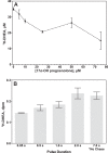
Pulse–chase assays of conversion of pregnenolone to DHEA. A, steady-state reaction. A reaction was initiated by mixing P450 17A1 (0.5 μm), NADPH-P450 reductase (2 μm), b5 (0.5 μm), DLPC (16 μm), and [3H]pregnenolone (13 μCi/μmol, in 50 mm potassium phosphate buffer) with an NADPH-generating system (68). After 60 s, the indicated concentration of unlabeled 17α-OH pregnenolone was added, and the reaction was continued for another 10 min, at which time the reaction was quenched by the addition of HCl (0.67 m, final), and the radiolabeled DHEA was measured by radio-HPLC as described under “Experimental procedures.” Results are presented as means ± S.D. (range) of duplicate determinations. B, rapid quench pulse–chase experiment. A similar approach was used, utilizing the rapid-quench apparatus with [3H]pregnenolone having a specific radioactivity of 830 μCi/μmol. The radiolabeled DHEA was measured after the addition of 80 μm unlabeled 17α-OH pregnenolone at the indicated times following initiation of the reaction with NADPH. The results are presented as means ± S.D. (range) of two individual experiments.
Figure 8.
Figure 8.
Inhibition of P450 17A1 reactions by (R)- and (S)-orteronel. Steady-state reactions were run with 0.01 μm P450 17A1 (0.1 μm for 17α-OH progesterone), in the presence of 0.5 μm b5, for 5 min in the presence of the indicated concentrations of the resolved enantiomers of orteronel. Results are presented as means of two determinations ± range, with fitting as described under “Experimental procedures.”
Figure 9.
Figure 9.
Kinetic scheme for oxidations of pregnenolone (Preg) and progesterone (Prog) using optimized rate constants and dissociation constants. See Fig. 10 for kinetic and binding data.
Figure 10.
Figure 10.
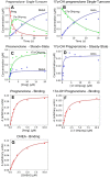
Fitting of pregnenolone kinetic and binding data with rate constants and Kd values (from Fig. 9). A and B are from Fig. 5, G and H, respectively (single-turnover results). C and D are steady-state kinetic results from Fig. 2, A and B, respectively. A, single-turnover kinetics beginning with pregnenolone (preg). B, single-turnover kinetics beginning with 17α-OH pregnenolone. C, steady-state kinetics for conversion of pregnenolone to 17α-OH pregnenolone and DHEA. D, steady-state kinetics of 17α-OH progesterone oxidation to DHEA. E, steady-state binding of pregnenolone to (ferric) P450 17A1. F, steady-state binding of 17α-OH pregnenolone to (ferric) P450 17A1. G, steady-state binding of DHEA to (ferric) P450 17A1.
Figure 11.
Figure 11.
Results from KinTek Explorer® data analysis with experimental Kd values as constants.
Figure 12.
Figure 12.
Analysis of P450 17A1 steroid binding kinetics. P450 17A1 (2 μm) was mixed with increasing concentrations of pregnenolone, 17α-OH pregnenolone, or DHEA as described in general under “Experimental procedures” (see Fig. 3 for concentrations, corresponding to the data points there; note, b5 was not present). Stopped-flow changes are shown in arbitrary units in the KinTek Explorer® global analysis, corresponding to ΔAaAb changes. A, pregnenolone; B, 17α-OH pregnenolone; C, DHEA. In the model, there are two forms of the enzyme, E and E*, and only one binds the ligand (to form EL). EL is in equilibrium with a second ligand-bound form, E′L. In the fitting, both EL and E′L show the observed spectral perturbation (and are therefore circled). Raw data are presented in the rough traces, and the overlaid lines are the fits from the KinTek Explorer® software. The wavelengths used for a and b changes were 391 and 426 for pregnenolone, 393 and 426 for 17α-OH pregnenolone, and 393 and 428 for DHEA. The values used were k1 = 0.23 s−1 and k−1 = 0.21 s−1 (for all cases) and the following for each ligand: pregnenolone, k2 = 1.2 × 10⁷ m−1 s−1; k−2 = 4.4 s−1; k3 = 0.50 s−1; and k−3 = 1.9 s−1; 17α-OH pregnenolone, k2 = 8.4 × 10⁶ m−1 s−1; k−2 = 2.2 s−1; k3 = 0.093 s−1; and k−3 = 2.3 s−1; DHEA, k2 = 1.2 × 10⁷ m−1 s−1; k−2 = 12 s−1; k3 = 0.10 s−1; and k−3 = 2.3 s−1.
Figure 13.
Figure 13.
Hypothetical P450 17A1 reaction scheme with additional conformations of ligand-bound enzyme. Δ5 steroids are shown, but the model can also be considered for Δ4 steroids.

Similar articles

Cited by

References

    1. Rendic S., and Guengerich F. P. (2015) Survey of human oxidoreductases and cytochrome P450 enzymes involved in the metabolism of xenobiotic and natural chemicals. Chem. Res. Toxicol. 28, 38–42 - PMC - PubMed
    1. Auchus R. J., and Miller W. L. (2015) in Cytochrome P450: Structure, Mechanism, and Biochemistry (Ortiz de Montellano P. R., ed) 4th Ed., pp. 851–879, Springer, New York
    1. Guengerich F. P. (2015) in Cytochrome P450: Structure, Mechanism, and Biochemistry (Ortiz de Montellano P. R., ed) 4th Ed., pp. 523–785, Springer, New York
    1. Auchus R. J. (2017) Steroid 17-hydroxylase and 17,20-lyase deficiencies, genetic and pharmacologic. J. Steroid Biochem. Mol. Biol. 165, 71–78 - PMC - PubMed
    1. Guengerich F. P. (2017) Intersection of roles of cytochrome P450 enzymes with xenobiotic and endogenous substrates. Relevance to toxicity and drug interactions. Chem. Res. Toxicol. 30, 2–12 - PMC - PubMed

Publication types

MeSH terms

LinkOut - more resources