. 2017 Jul 7;357(6346):eaal3753.
doi: 10.1126/science.aal3753.

Multipotent peripheral glial cells generate neuroendocrine cells of the adrenal medulla

Affiliations

Multipotent peripheral glial cells generate neuroendocrine cells of the adrenal medulla

Alessandro Furlan et al. Science. .

Abstract

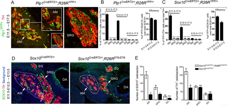
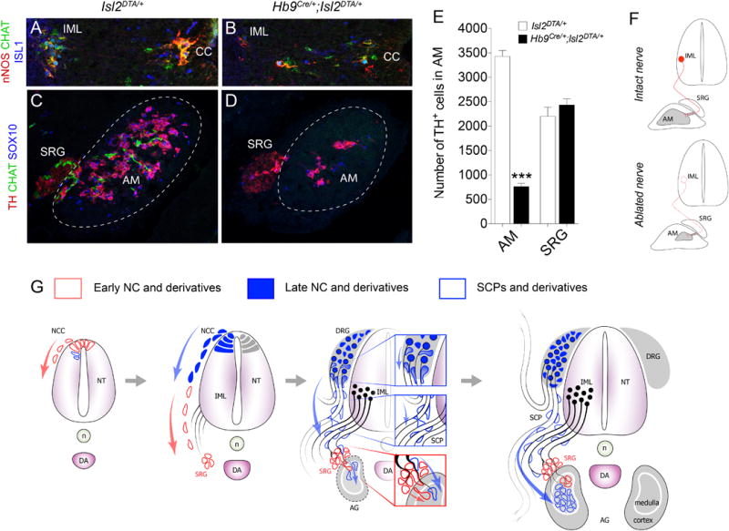
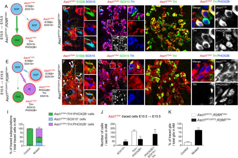
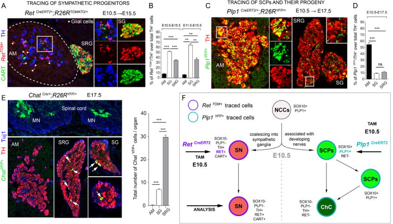
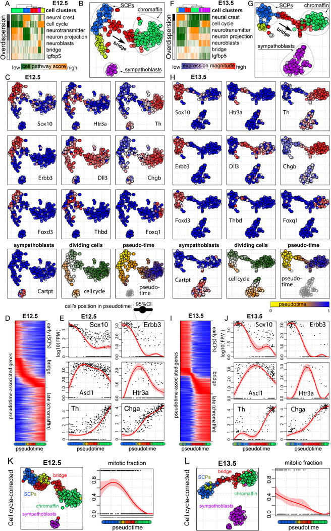
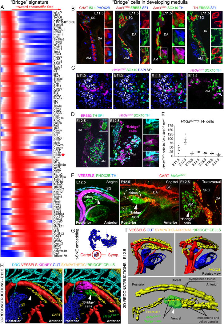
Adrenaline is a fundamental circulating hormone for bodily responses to internal and external stressors. Chromaffin cells of the adrenal medulla (AM) represent the main neuroendocrine adrenergic component and are believed to differentiate from neural crest cells. We demonstrate that large numbers of chromaffin cells arise from peripheral glial stem cells, termed Schwann cell precursors (SCPs). SCPs migrate along the visceral motor nerve to the vicinity of the forming adrenal gland, where they detach from the nerve and form postsynaptic neuroendocrine chromaffin cells. An intricate molecular logic drives two sequential phases of gene expression, one unique for a distinct transient cellular state and another for cell type specification. Subsequently, these programs down-regulate SCP-gene and up-regulate chromaffin cell-gene networks. The AM forms through limited cell expansion and requires the recruitment of numerous SCPs. Thus, peripheral nerves serve as a stem cell niche for neuroendocrine system development.


Figures

Fig. 1
Fig. 1. Chromaffin cells of the adrenal gland originate from PLP1+ and SOX10+ Schwann cell precursors (SCPs) at E11.5 and E12.5
A. Immunohistochemistry for YFP (recapitulating Plp1 expression) and TH on sections of the developing adrenal medulla (AM) and suprarenal ganglion (SRG) following genetic tracing in Plp1CreERT2/+;R26RYFP/+ animals injected with TAM at E11.5 and analyzed at E17.5. The arrow points at the developing AM. B-C. Quantification of the proportion of TH+/Plp1YFP+ cells traced in Plp1CreERT2/+;R26RYFP/+ (B) and of TH+/Sox10YFP+ cells in the AM, SRG and sympathetic ganglia (SG) of Sox10CreERT2/+;R26RYFP/+ mice (C) injected at E11.5, E12.5 or E15.5 and analyzed at E17.5. Note that the recombination efficiency (percentage of SOX10+/Plp1YFP+ cells out of all SOX10+ cells in embryonic nerves) at E12.5 is lower than that at E11.5. D. Immunohistochemistry for SOX10 and TH on E13.5 sections of developing AM and SRG following tamoxifen (TAM)-induced cell ablation of SOX10+ cells at both E11.5 and E12.5 in Sox10CreERT2/+;R26RDTA/DTA mice. Note the marked decrease in SOX10+ and TH+ cell numbers in Sox10CreERT2/+;R26RDTA/DTAembryos as compared to Sox10CreERT2/+;R26R+/+. E. Quantification of SOX10+ and TH+ cell numbers identified in the AM, SRG and SG of Sox10CreERT2/+;R26RDTA/DTA and Sox10CreERT2/+;R26R+/+embryos. Data are presented as mean ± s.e.m., two-tailed Student t-test. In A-C, for injection at E11.5: n=3, at E12.5: n=4 and at E15.5: n=3. In D-E, for Sox10CreERT2/+;R26R+/+ and Sox10CreERT2/+;R26RDTA/DTA AM n=3, SRG n=4, SG n=3. AM: adrenal medulla; SRG: suprarenal ganglion; SG: sympathetic ganglion; ns: non-significant.
Fig. 2
Fig. 2. Pre-ganglionic nerves are necessary for adrenal medulla assembly
A,B Immunohistochemistry for nNOS, CHAT and ISL1 on E14.5 sections of spinal cords from control Isl2DTA/+ (A) and Hb9Cre/+;Isl2DTA/+ embryos (B) show almost complete ablation of CHAT+/nNOS+/ISL1+ pre-ganglionic neurons. C, D. Immunohistochemistry for TH, SOX10 and CHAT on E14.5 sections of adrenal medulla (AM) from control Isl2DTA/+ (C) and nerve-ablated Hb9Cre/+;Isl2DTA/+ embryos (D). Note the significant reduction of chromaffin cells in the AM, but not of suprarenal ganglion (SRG) sympathetic neurons in nerve-ablated (D) compared to control (C) embryos. (E). Quantification of C, D. Data are presented as mean ± s.e.m., n=3 for Isl2DTA/+, n=5 for Hb9Cre/+;Isl2DTA/+, two-tailed Student t-test. F. Graphical summary of the results. G. Schematic showing the origin of chromaffin cells from nerve-associated SCPs. IML: intermediolateral cell column; CC: central canal; AM: adrenal medulla; AG: adrenal gland; SRG: suprarenal ganglion; NCC: neural crest cells; NC: neural crest; NT: neural tube; n: notochord; DA: dorsal aorta; DRG: dorsal root ganglion; SCPs: Schwann cell precursors.
Figure 3
Figure 3. The role of Ascl1 in the SCP-to-chromaffin transition
A-H Genetic ablation of Ascl1 prevents the glia-to-chromaffin transition. The majority of nerve-associated SOX10+/S100β+ cells expressing Ascl1 at E10.5 downregulated glial markers in Ascl1CreERT2/+;R26RTOM/+ (control) embryos at E15.5 and have differentiated towards TH+/PHOX2B+ chromaffin cells (A,B,C) but failed to do so in Ascl1-deficient Ascl1CreERT2/CreERT2;R26RTOM/+ (mutant) embryos (E,F,G), while a new, intermediate PHOX2B+/TH/SOX10 population is observed (D,H). I,J. The majority of Ascl1TOM+ cells in Ascl1CreERT2/+;R26RTOM/+ embryos are chromaffin cells with a minor contribution to SOX10+ glia, while in the Ascl1-deficient Ascl1CreERT2/CreERT2;R26RTOM/+ embryos the larger population is represented by PHOX2B+/TH/SOX10 cells with simultaneous increase in SOX10+ glia and reduction in TH+/PHOX2B+ chromaffin cells. K. The proportion of Ascl1TOM+/SOX10+ glia over the total glia population is significantly increased in the mutant. Note the complete absence of the Ascl1TOM+-traced PHOX2B+/TH/SOX10 population in the control, which is observed in the mutant. Data presented as mean ± s.e.m., n=3 for all cases, two-tailed Student t-test. In A and E, the main differentiation trajectories derived from Schwann cell precursors (SCPs – marked with a pink halo) are shown with big arrows, while trajectories producing a minor or reduced cell population are shown by dashed arrows. White arrowheads point to Ascl1TOM+ glial cells. SCP: Schwann cell precursor; ChC: chromaffin cell; IC: intermediate cell; AM: adrenal medulla.
Figure 4
Figure 4. Early specification and separation of sympathoadrenal lineages during development
A. Immunohistochemistry for TOMATO (recapitulating Ret expression) on RetCreERT2/+;R26RTOM/+embryos injected with TAM at E10.5 and analysed at E15.5. Arrowheads point to traced glia. B. Analysis of Ret tracing in the adrenal medulla (AM), sympathetic ganglion (SG) and suprarenal ganglion (SRG) in RetCreERT2/+;R26RTOM/+embryos injected with TAM at E10.5 or E11.5 and analysed at E15.5. Note the high recombination in the SG and SRG (for E10.5 injection: 51.77±1.40% in the SG, 35.05±1.19% in the SRG; for E11.5 injection: 64.96±3.01% in the SG, 36.4±3.08% in the SRG) in contrast to the AM (for E10.5 injection: 8.09±0.71%; for E11.5 injection: 6.54±0.72%). C. Immunohistochemistry for YFP (recapitulating Plp1 expression) and TH on Plp1CreERT2/+/R26RYFP/+ embryos injected with TAM at E10.5 and analyzed at E17.5. Note high numbers of Plp1YFP+/TH+ cells in the AM but only a few in the SG and SRG. D. Quantification of C. Note the high recombination in the AM (54.60±3.01%) in contrast to the SG and SRG (9.23±0.22% and 11.05±1.37% respectively). E. Immunohistochemistry for YFP (recapitulating Chat expression), TH and TuJ1 on ChatCre/+;R26RYFP/+ embryos analyzed at E17.5. Arrows point to ChatYFP+ cells in the SRG and SG. F. Schematic showing the fate restriction of neural crest cells towards sympathetic neurons and Schwann cell precursors, finally differentiating into chromaffin cells. In B,D,E data are presented as mean ± s.e.m., n=3 for all cases, two-tailed Student t-test. SG: sympathetic ganglion; SRG: suprarenal ganglion; AM: adrenal medulla; DRG: dorsal root ganglion; MN: motoneurons; TAM: tamoxifen; NCCs: neural crest cells; SN: sympathetic neurons; SCPs: Schwann cell precursors; ChC: chromaffin cell.
Fig. 5
Fig. 5. Transcriptional heterogeneity of the developing adrenal medulla demonstrates SCP-to-chromaffin fate transition through a defined “bridge” state
A. Distinct subpopulations of neural crest-derived cells (columns) are seen within the E12.5 adrenal medulla (AM). Top 5 clusters of cells are given by the color bar below the dendrogram. Top 7 statistically significant aspects (rows) of transcriptional heterogeneity are shown, labelled according to the main GO category or a key gene driving each aspect. B. Transcriptional profile-based subpopulations in the E12.5 sample are visualized using t-SNE embedding. The labels show interpretation of the distinct subpopulations based on the key marker genes below, and a continuous “bridge” cell population (in red) connecting the SCPs and the chromaffin clusters. C. Expression levels of representative marker genes within the E12.5 population (first 10 plots). Cell cycle dynamics (“dividing cells”: yellow – mitotic, green – interphase) are also shown. Finally, position of each cell along the SCP-to-chromaffin differentiation trajectory pseudotime is shown in the last plot. D. Expression profiles of 1480 genes (rows) that are statistically significantly associated with the differentiation trajectory are shown as a function of pseudotime (cell position, x axis; individual cells colored according to their cluster membership are shown right below the axis). The genes can be categorized into those expressed early (close to SCPs), late (close to chromaffin) and transiently (within the”bridge”subpopulation). E. Examples of pseudotime expression profiles for key genes within each category. Each point represents a cell, with the small horizontal line around it indicating uncertainty (95% CI) of the cell’s position within the differentiation pseudotime. A smoothed regression line with the associated 95% CI is shown in red. F-J. Analogous subpopulation and SCP-to-chromaffin differentiation”bridge”can be seen in the neural crest-derived cells of the E13.5 adrenal medulla. In panel A, the greyscale gradient represents z-scores from 4.3 to 63, while in panel F from 5 to 105. K, L. t-SNE plots of developing medulla subpopulations normalized for the cell cycle-correlated genes at E12.5 (K) and E13.5 (L). Note that yellow population aligns with the red “bridge” population. The graphs show the fraction of cells undergoing mitosis (y-axis) as a function of pseudotime. Cells were classified as mitotic in the case of a positive score on the mitosis-driven aspect of heterogeneity.
Fig. 6
Fig. 6. Mapping a novel “bridge” population of cells to the anatomical location and developmental timeline
A. Genes in the “bridge” cell population whose expression is dynamically changing in a statistically significant way. B. Left: immunohistochemistry for CHAT, PHOX2B and ISL1 on sections of adrenal medulla (AM) region from E11.5 embryos. Note that PHOX2B+ cells are associated with CHAT+ nerve. Center: in 24h-traced Ascl1CreERT2/+;R26RTOM/TOM embryos analyzed at E11.5, nerve-associated cells of the intermediate stage express Ascl1 in addition to the glial markers ERBB3 and SOX10. Note that TH+/Ascl1TOM+ cells retain low levels of SOX10. Right: immunohistochemistry at E11.5 for TH, ERBB3 and SF1 shows that TH+ cells near the assembling adrenal cortex (SF1+ cells) are associated with the nerve but do not express glial markers. C-D. Validation of the “bridge” cell population in SCP-to-chromaffin transition marked by the expression of Htr3aEGFP in E11.5 (C) and E12.5 (D) embryos. In C, immunohistochemistry for GFP (recapitulating Htr3a expression), SOX10 and SF1 shows the anatomical position of the very first Htr3aEGFP AM cells within the developing adrenal gland region (marked by SF1 expression). Right: immunohistochemistry for GFP (recapitulating Htr3a expression), SOX10 and TH shows that intermediate “bridge” Htr3aEGFP cells of AM rarely express TH but retained variable levels of SOX10. In D, AM cells are either intermediate “bridge” TH+/Htr3aEGFPlow and SOX10+/Htr3aEGFPlow or chromaffin TH+/Htr3aEGFP− cells. E. Htr3aEGFP+ cells number in the medulla region peaks at E12.5, declines at E13.5 and keeps constant throughout development. Each dot represents counts from one medullary region (n=2 embryos for all ages). F. Left panel: whole mount immunofluorescence for PHOX2B and TH (magenta: auto-fluorescent blood vessels), CART and Htr3aEGFP (middle panel) and immunohistochemistry on transversal cryosection (right panel). G. Note that Cartpt (gene coding for CART) is not expressed in the intermediate Htr3aEGFP+ “bridge” cells or differentiating chromaffin cells, while it is present in sympathetic neurons of the SRG. H, I. Corresponding 3D-reconstructions of sympatho-adrenal structures of E12.5 embryos. Large blood vessels were reconstructed based on blood vessel auto-fluorescence of embryonic erythrocytes (see F). Sympathetic structures were reconstructed based on CART+ and PHOX2B+ signal. Note the consolidation of ventral sympathetic structures (presumably SRG, para-aortic, future mesenteric and celiac ganglia) at this stage as well as the presence of the intermediate “bridge” cells outlined in the AM by Htr3aEGFP signal (white arrows). SG: sympathetic ganglion; DA: dorsal aorta; AM: adrenal medulla; SRG: suprarenal ganglion.

Similar articles

Cited by

References

    1. Huber K, Kalcheim C, Unsicker K. The development of the chromaffin cell lineage from the neural crest. Auton Neurosci. 2009;151:10–16. - PubMed
    1. Saito D, Takase Y, Murai H, Takahashi Y. The dorsal aorta initiates a molecular cascade that instructs sympatho-adrenal specification. Science. 2012;336:1578–1581. - PubMed
    1. Ernsberger U, et al. Expression of neuronal markers suggests heterogeneity of chick sympathoadrenal cells prior to invasion of the adrenal anlagen. Cell Tissue Res. 2005;319:1–13. - PubMed
    1. Chan WH, et al. Differences in CART expression and cell cycle behavior discriminate sympathetic neuroblast from chromaffin cell lineages in mouse sympathoadrenal cells. Dev Neurobiol. 2016;76:137–149. - PMC - PubMed
    1. Gut P, et al. Lack of an adrenal cortex in Sf1 mutant mice is compatible with the generation and differentiation of chromaffin cells. Development. 2005;132:4611–4619. - PubMed

Publication types

MeSH terms