. 2017 Jul 7;28(14):1833-1846.
doi: 10.1091/mbc.E17-03-0134.

Single and collective cell migration: the mechanics of adhesions

Affiliations

Single and collective cell migration: the mechanics of adhesions

Chiara De Pascalis et al. Mol Biol Cell. .

Abstract

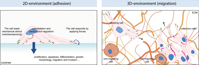
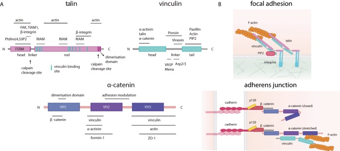
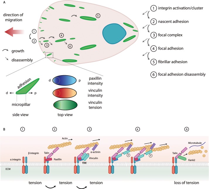
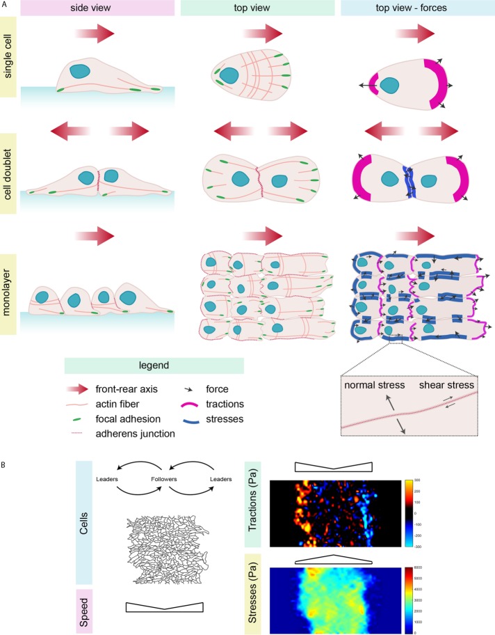
Chemical and physical properties of the environment control cell proliferation, differentiation, or apoptosis in the long term. However, to be able to move and migrate through a complex three-dimensional environment, cells must quickly adapt in the short term to the physical properties of their surroundings. Interactions with the extracellular matrix (ECM) occur through focal adhesions or hemidesmosomes via the engagement of integrins with fibrillar ECM proteins. Cells also interact with their neighbors, and this involves various types of intercellular adhesive structures such as tight junctions, cadherin-based adherens junctions, and desmosomes. Mechanobiology studies have shown that cell-ECM and cell-cell adhesions participate in mechanosensing to transduce mechanical cues into biochemical signals and conversely are responsible for the transmission of intracellular forces to the extracellular environment. As they migrate, cells use these adhesive structures to probe their surroundings, adapt their mechanical properties, and exert the appropriate forces required for their movements. The focus of this review is to give an overview of recent developments showing the bidirectional relationship between the physical properties of the environment and the cell mechanical responses during single and collective cell migration.


Figures

FIGURE 1:
FIGURE 1:
Mechanobiology and migration. Schematic of cells migrating on two-dimensional (2D) or 3D matrices. The 2D example shows the principles of mechanobiology by which a cell reads (green arrows) the mechanical properties of the ECM and converts them into a biochemical intracellular signal (red arrow) that affects the cytoskeleton, signaling, and transcription. Ultimately, the cell responds both by applying forces to the matrix itself (blue arrow) and undergoing processes such as proliferation, apoptosis, differentiation, and migration (large blue arrow). Mechanosensing occurs as the cells interact with the ECM through focal adhesions (green), which are linked to actin fibers (pink). The situation is more complicated in 3D migration, in which cells can move inside a matrix, here composed of fibers (different shades of orange) of different composition, structure, topology, and rigidity and other nonmigrating cells (pink). The drawing shows both a single invading cell (pink), moving in the direction of the arrow, and a group of migrating cells (pink) moving collectively and attached to one another by cell–cell junctions (magenta). Attached to the group of cells there can also be nonmigrating, nonpolarized cells (brown). In this complex situation, cells must integrate the signals transmitted by different types of focal adhesions and adherens junctions.
FIGURE 2:
FIGURE 2:
Tension-sensitive proteins are mechanical players of adhesion sites. (A) Schematic representation of the main tension-sensitive proteins involved in focal adhesions and adherens junctions: talin (purple), vinculin (light blue), and α-catenin (dark blue). The main protein interaction domains are shown, and the known interactors (with their binding sites) are indicated above or below each protein. (B) Top, components of focal adhesions and the structures of talin and vinculin when stretched (talin in pink, vinculin in light blue, PIP2 in purple, and F-actin in orange). Bottom, components of adherens junctions and the structure of the unstretched (closed, top) or stretched (bottom) α-catenin (cadherin in purple, p120 in yellow, β-catenin in coral, α-catenin in purple, vinculin in light blue, and actin in orange).
FIGURE 3:
FIGURE 3:
Turnover of focal adhesions. (A) Schematic representation of a migrating cell (pink; nucleus in blue; the arrow shows the direction of migration), highlighting focal adhesion (green) formation, maturation, and disassembly. Different maturation stages (labeled 1–6) can be observed, which can grow (full arrow) or disassemble (dotted arrow) at every step. Inset, summary of the work by Sarangi et al. (2016). The intensity of vinculin and paxillin is analyzed in parallel to vinculin tension (green, high; to white, low) on micropillars. The intensity of paxillin (blue, high; to white, low) and vinculin (red, high; to white, low) is higher in the region of the focal adhesion corresponding to the edge of the micropillar (yellow dotted lines), whereas the vinculin tension is higher at the distal (d) and proximal (p) sites in the adhesion. (B) Focal adhesions, from an integrin cluster to a mature focal adhesion that forms with tension. The disassembly occurs with loss of tension. The ECM (green), integrins (green and red), paxillin (purple), talin (pink), vinculin (light blue), FAK (blue), α-actinin (purple), actin (yellow), microtubules (blue line), and Kank2 (green).
FIGURE 4:
FIGURE 4:
Cell migration and force transmission and their study in collective migration. (A) Single cell (top), doublets (middle), and a migrating monolayer (bottom) from the side and top views. Cells (light pink) show a polarized (red arrow, front–rear axis of migration) morphology, with the nucleus (blue) at the back, an asymmetric distribution of focal adhesions (green) and actin (pink lines), microtubule (not shown), and intermediate filament (not shown) networks. Cell–cell contacts (red dotted line) allow adhesion between cells. The third column shows representative forces (tractions on the substrate in magenta and intercellular stresses in blue); tractions are high at the cell front, whereas intercellular stresses concentrate at cell–cell contacts. The gray arrows represent possible forces and their directions. (B) Representative images of cells migrating on hydrogels (black lines show the cell edges). Collectively migrating cells are divided into leaders and followers, which influence one another. The speed of migration is higher at the edges of the monolayer, as are tractions. Tractions are calculated by TFM (bead displacements) and are higher where the color intensity is stronger—yellow and blue correspond to the maximal forces in opposite directions along the axis of migration. Stresses, calculated with MSM, are higher at the center of the migrating monolayer (strong intensity in red). TFM and MSM images were obtained on migrating astrocytes on a 9-kPa collagen-coated hydrogel by C.D.P. and C. Pérez González.

References

    1. Akhmanova A, Stehbens SJ, Yap AS. Touch, grasp, deliver and control: functional cross-talk between microtubules and cell adhesions. Traffic. 2009;10:268–274. - PubMed
    1. Aleksandrova A, Czirok A, Kosa E, Galkin O, Cheuvront TJ, Rongish BJ. The endoderm and myocardium join forces to drive early heart tube assembly. Dev Biol. 2015;404:40–54. - PMC - PubMed
    1. Aragona M, Panciera T, Manfrin A, Giulitti S, Michielin F, Elvassore N, Dupont S, Piccolo S. A mechanical checkpoint controls multicellular growth through YAP/TAZ regulation by actin-processing factors. Cell. 2013;154:1047–1059. - PubMed
    1. Aranjuez G, Burtscher A, Sawant K, Majumder P, McDonald JA. Dynamic myosin activation promotes collective morphology and migration by locally balancing oppositional forces from surrounding tissue. Mol Biol Cell. 2016;27:1898–1910. - PMC - PubMed
    1. Atherton P, Stutchbury B, Jethwa D, Ballestrem C. Mechanosensitive components of integrin adhesions: role of vinculin. Exp Cell Res. 2016;343:21–27. - PMC - PubMed

LinkOut - more resources