. 2017 Sep;37(9):1646-1656.
doi: 10.1161/ATVBAHA.117.309510. Epub 2017 Jul 6.

Endothelial Cell Autophagy Maintains Shear Stress-Induced Nitric Oxide Generation via Glycolysis-Dependent Purinergic Signaling to Endothelial Nitric Oxide Synthase

Affiliations

Endothelial Cell Autophagy Maintains Shear Stress-Induced Nitric Oxide Generation via Glycolysis-Dependent Purinergic Signaling to Endothelial Nitric Oxide Synthase

Leena P Bharath et al. Arterioscler Thromb Vasc Biol. 2017 Sep.

Abstract

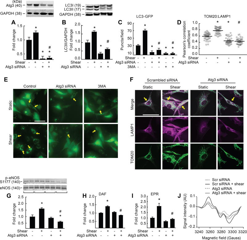
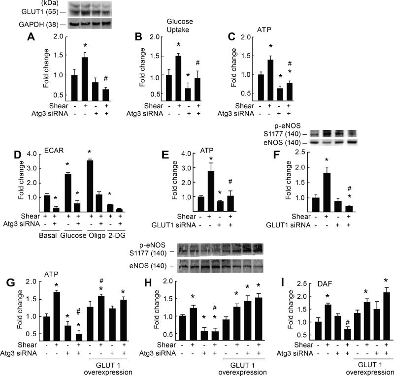
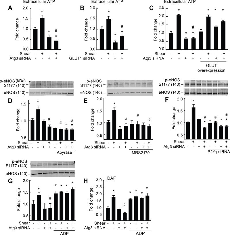
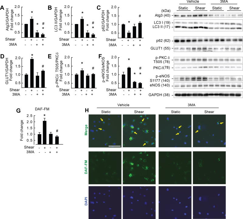
Objective: Impaired endothelial cell (EC) autophagy compromises shear stress-induced nitric oxide (NO) generation. We determined the responsible mechanism.

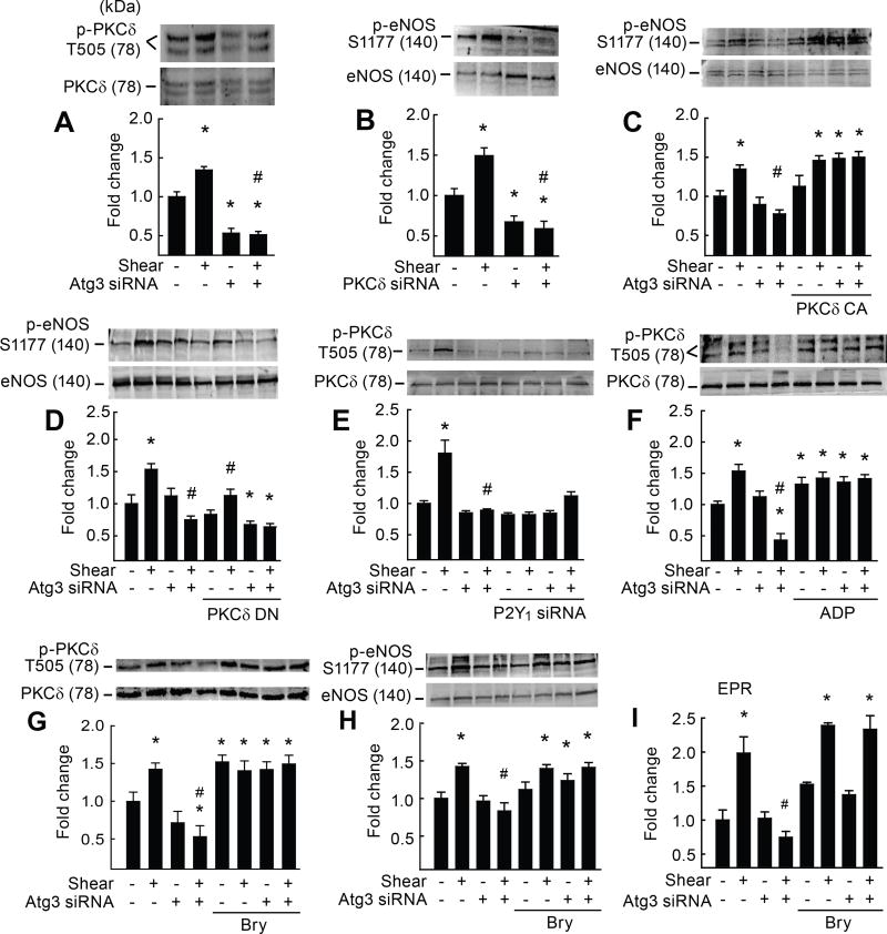
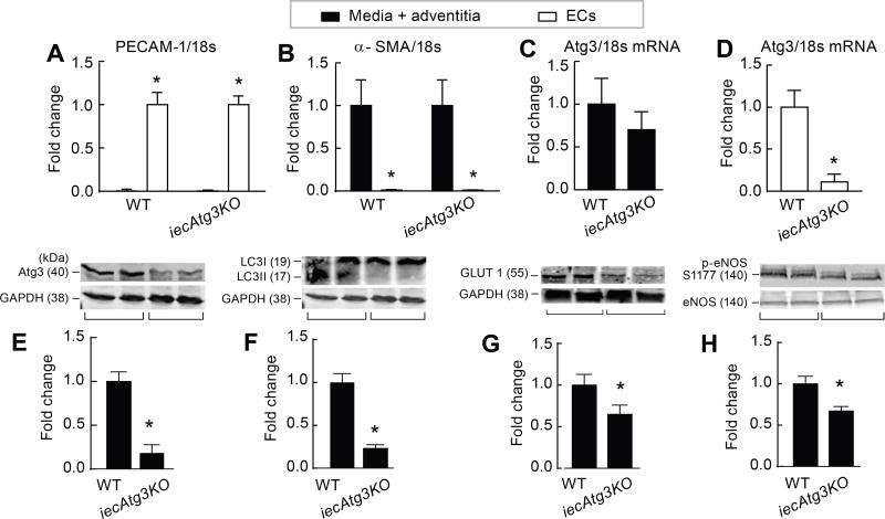
Approach and results: On autophagy compromise in bovine aortic ECs exposed to shear stress, a decrease in glucose uptake and EC glycolysis attenuated ATP production. We hypothesized that decreased glycolysis-dependent purinergic signaling via P2Y1 (P2Y purinoceptor 1) receptors, secondary to impaired autophagy in ECs, prevents shear-induced phosphorylation of eNOS (endothelial nitric oxide synthase) at its positive regulatory site S1117 (p-eNOSS1177) and NO generation. Maneuvers that restore glucose transport and glycolysis (eg, overexpression of GLUT1 [glucose transporter 1]) or purinergic signaling (eg, addition of exogenous ADP) rescue shear-induced p-eNOSS1177 and NO production in ECs with impaired autophagy. Conversely, inhibiting glucose transport via GLUT1 small interfering RNA, blocking purinergic signaling via ectonucleotidase-mediated ATP/ADP degradation (eg, apyrase), or inhibiting P2Y1 receptors using pharmacological (eg, MRS2179 [2'-deoxy-N6-methyladenosine 3',5'-bisphosphate tetrasodium salt]) or genetic (eg, P2Y1-receptor small interfering RNA) procedures inhibit shear-induced p-eNOSS1177 and NO generation in ECs with intact autophagy. Supporting a central role for PKCδT505 (protein kinase C delta T505) in relaying the autophagy-dependent purinergic-mediated signal to eNOS, we find that (1) shear stress-induced activating phosphorylation of PKCδT505 is negated by inhibiting autophagy, (2) shear-induced p-eNOSS1177 and NO generation are restored in autophagy-impaired ECs via pharmacological (eg, bryostatin) or genetic (eg, constitutively active PKCδ) activation of PKCδT505, and (3) pharmacological (eg, rottlerin) and genetic (eg, PKCδ small interfering RNA) PKCδ inhibition prevents shear-induced p-eNOSS1177 and NO generation in ECs with intact autophagy. Key nodes of dysregulation in this pathway on autophagy compromise were revealed in human arterial ECs.

Conclusions: Targeted reactivation of purinergic signaling and PKCδ has strategic potential to restore compromised NO generation in pathologies associated with suppressed EC autophagy.

Keywords: autophagy; cell physiological phenomena; endothelial cells; nitric oxide; reactive oxygen species.


Figures

Figure 1
Figure 1. Genetic disruption of Atg3 impairs shear-stress induced autophagy, mitophagy, and NO generation
Relative to static conditions, shear-stress increased Atg3 protein expression (A), LC3 II accumulation (B), LC3-GFP puncta formation (C, E), colocalization of TOM20 with LAMP1 (D, F), p-eNOSS1177 (G), and NO generation (H–J), in ECs transfected with scrambled siRNA (bar 1 vs. 2) but not in ECs transfected with Atg3 siRNA (bar 3 vs. 4), or in ECs after treatment with 3MA (C, bar 5 vs. 6). Images shown in E, F, and J represent mean data shown in C, D, and I, respectively. Fluorescence images in E, F were individually adjusted to maximize clarity. Calibration bar = 50 µm. For A, B (n=30), C, D (n=10), E, F (n=6), G, H (n=30), I, J (n=10). For A, B, G each n=1 × 10 cm petri dish. For C, D each n=1 well of a 24-well plate. For H, each n = 1 well of a 6-well plate. For C, D, E, F each n=10 cells per field × 10 fields per slide. For I, J each n = 3 wells of a 6-well plate. *p<0.05 vs. (−shear)(−Atg3 siRNA); # p<0.05 vs. (+shear)(−Atg3 siRNA).
Figure 2
Figure 2. Genetic disruption of Atg3 impairs EC glycolysis and NO generation
Relative to static conditions, shear-stress increased GLUT1 protein expression (A), glucose uptake (B), ATP production (C, G), p-eNOSS1177 (H) and NO generation (I) in ECs transfected with scrambled siRNA (bar 1 vs. 2) but not Atg3 siRNA (bar 3 vs. 4). For A–C (n=8–12). For A, each n = 1 × 10 cm petri dish). For B, C, each n = 1 well of a 6-well plate. For A–C *p<0.05 vs. (−shear)(−Atg3 siRNA); #p<0.05 vs. (+shear)(−Atg3 siRNA). Extracellular acidification rate (ECAR) was assessed in ECs exposed to shear stress ± Atg3 siRNA (D). Relative to basal conditions, ECs transfected with scrambled siRNA displayed increased ECAR in response to 5 mM glucose (bar 1 vs. 3) and 1 µM oligomycin (oligo; bar 1 vs. 5), but these responses were not observed in ECs after Atg3 siRNA (bar 2 vs. 4; bar 2 vs. 6, respectively). Relative to basal conditions, ECAR decreased upon treatment with 50 mM 2-DG in ECs transfected with scrambled (bar 1 vs. 7) but not Atg3 siRNA (bar 2 vs. 8). For D, n=3, each n= 1 seahorse plate. For D, *p<0.05 vs. basal condition (+shear)(−Atg3 siRNA i.e., bar 1); # p<0.05 vs. same condition (+shear)(−Atg3 siRNA). Relative to static conditions, shear-stress increased ATP (E) and p-eNOSS1177 (F) in ECs transfected with scrambled siRNA (bar 1 vs. 2) but not GLUT1 siRNA (bar 3 vs. 4). For E, n=12, each n = 1 well of a 6-well plate. For F, n=6, each n = 1 × 10 cm petri dish. For E, F *p<0.05 vs. (−shear)(−Atg3 siRNA); #p<0.05 vs. (+shear)(−Atg3 siRNA). After co-transfection with a plasmid vector to increase GLUT1 expression in ECs (G–I), the suppression of shear-induced ATP (G), p-eNOSS1177 (H), and NO generation (I) after autophagy repression (bar 2 vs. 4) was not observed (bar 6 vs. 8). For G–I, n=4, each n = 1 × 10 cm petri dish). For G–I *p<0.05 vs. (−shear)(−Atg3 siRNA); #p<0.05 vs. (+shear)(−Atg3 siRNA).
Figure 3
Figure 3. Genetic disruption of Atg3 limits purinergic-mediated activation of eNOS
Relative to static conditions, shear-stress increased extracellular ATP accumulation (A–C), p-eNOSS1177 (D–G), and NO generation (H) in ECs transfected with scrambled siRNA (bar 1 vs. 2) but not Atg3 siRNA (A,C,D,E; bar 3 vs. 4) or GLUT1 siRNA (B; bar 3 vs. 4). After co-transfection with a plasmid vector to increase GLUT1 expression in ECs (C), the suppression of shear-induced ATP after autophagy compromise (bar 2 vs. 4) was normalized (bar 6 vs. 8). The ectonucleotidase apyrase (D), the pharmacological P2Y1-R blocker MRS2179 (E), and genetic disruption of P2Y1-R via siRNA (F) prevented shear-induced p-eNOSS1177 in ECs with intact Atg3 protein (bar 5 vs. 6). Conversely exogenous 2-methylthioadenosine diphosphate (ADP) restored shear-induced p-eNOSS1177 (G) and NO generation (H) in ECs with suppressed autophagy (bar 7 vs. 8). For A–C, H, n=6, each n = 1 well of a 6-well plate. For D–G, n=5, each n = 1 × 10 cm petri dish. * p<0.05 vs. (−shear)(−Atg3 siRNA); # p<0.05 vs. (+shear)(−Atg3 siRNA).
Figure 4
Figure 4. Defective purinergic-mediated p-PKCδT505 activation of eNOS after autophagy compromise is normalized by genetic and pharmacological approaches
Relative to static conditions, shear-stress increased p-PKCδT505 (A,E,F,G) and p-eNOSS1177 (C,D,H) in ECs transfected with scrambled siRNA (bar 1 vs. 2) but not Atg3 siRNA (bar 3 vs. 4). Relative to static conditions, shear-stress increased p-eNOSS1177 in ECs transfected with scrambled but not PKCδ siRNA (B) (bar 2 vs. 4). Suppressed shear-induced p-eNOSS1177 after Atg3 siRNA was restored in ECs co-transfected with constitutively active (CA) PKCδ (bar 7 vs. 8; C) but not dominant-negative (DN) PKCδ (D). Suppressed shear-induced p-PKCδT505 after Atg3 siRNA (A,E,F,G) could be recapitulated in ECs with intact Atg3 protein that were transfected with P2Y1-R siRNA (bar 5 vs. 6; E) or restored in ECs with autophagy compromise by ADP (bar 7 vs. 8; F). Suppressed shear-induced p-PKCδT505, p-eNOSS1177, and NO generation in ECs transfected with Atg3 siRNA was restored by the PKCδ agonist bryostatin-1 (bry; bar 4 vs. 8, G, H, I). For A–H, n=5, each n = 1 × 10 cm petri dish. For I, n=6, each n = 3 wells of a 6-well plate. *p<0.05 vs. (−shear)(−Atg3 siRNA); # p<0.05 vs. (+shear)(−Atg3 siRNA).
Figure 5
Figure 5. Pharmacological inhibition of autophagy impairs shear-stress induced NO generation in human arterial endothelial cells
Relative to static conditions, shear-stress increased Atg3 (A), LC3 II (B), GLUT1 (D), p-PKCδT505 (E) and p-eNOSS1177 (F) protein expression, NO generation (G), and p62 degradation (C) (bar 1 vs. 2). All responses were prevented by concurrent treatment with 3MA (bar 3 vs. 4). Images shown in the “merge” portion of H represent mean data shown in G. Calibration bar = 100 µm. For A–F, n=3, each n= 2 wells of a 6-well plate. For G and H, n=2, each n = 1 well of a 4 well chamber slide. *p<0.05 vs. (−shear)(− 3MA); # p<0.05 vs. (+shear)(− 3MA).
Figure 6
Figure 6. iecAtg3KO mice exhibit impaired endothelial cell autophagy, GLUT1 protein expression, and p-eNOS protein expression vs. WT mice
To assess mRNA, endothelial cells (ECs) and media + adventitia were isolated from iliac arteries of 5-month old iecAtg3KO and WT mice treated 30-days earlier with tamoxifen. Purity of the EC and media + adventitia fraction was confirmed by PECAM (A) and α-SMA (B) staining, respectively. Atg3/18S mRNA was similar in the media + adventitia component of both groups (C), but was lower in the EC fraction of iecAtg3KO vs. WT mice (D). For A–D, n=4 mice per group. *p<0.05 vs. media + adventitia (A); vs. EC (B); vs. WT (D). EC protein was assessed in a different cohort of 5-month old mice treated 30 days earlier with tamoxifen. Atg3:GAPDH (E), LC3 II:GAPDH (F), GLUT1 : GAPDH (G), and p-eNOS : eNOS (H) was lower in ECs from iecAtg3KO vs. WT mice. For E–H, n=3, each n = ECs obtained from 4 entire aortae. *p<0.05 vs. WT.

References

    1. Zhang Y, Janssens SP, Wingler K, Schmidt HH, Moens AL. Modulating endothelial nitric oxide synthase: a new cardiovascular therapeutic strategy. Am J Physiol Heart Circ. Physiol. 2011;301:H634–H646. - PubMed
    1. Triggle CR, Hollenberg M, Anderson TJ, Ding H, Jiang Y, Ceroni L, Wiehler WB, Ng ESM, Ellis A, Andrews K, McGuire JJ, Pannirselvam M. The endothelium in health and disease- a target for therapeutic intervention. J Sm Musc. 2003;39:249–267. - PubMed
    1. Kuma A, Mizushima N. Physiological role of autophagy as an intracellular recycling system: with an emphasis on nutrient metabolism. Semin Cell Dev Biol. 2010;21:683–690. - PubMed
    1. Mizushima N. Physiological functions of autophagy. Curr Top Microbiol Immunol. 2009;335:71–84. - PubMed
    1. Larocca TJ, Henson GD, Thorburn A, Sindler AL, Pierce GL, Seals DR. Translational evidence that impaired autophagy contributes to arterial ageing. J Physiol. 2012;590:3305–3316. - PMC - PubMed

Publication types

MeSH terms