. 2017 Jul 6;7(1):4802.
doi: 10.1038/s41598-017-05001-z.

MiR-320a as a Potential Novel Circulating Biomarker of Arrhythmogenic CardioMyopathy

Affiliations

MiR-320a as a Potential Novel Circulating Biomarker of Arrhythmogenic CardioMyopathy

Elena Sommariva et al. Sci Rep. .

Abstract

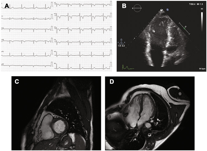
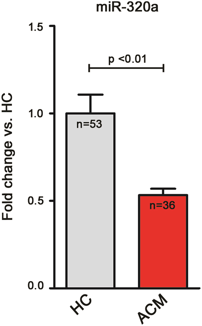
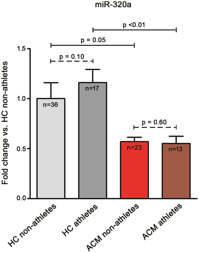
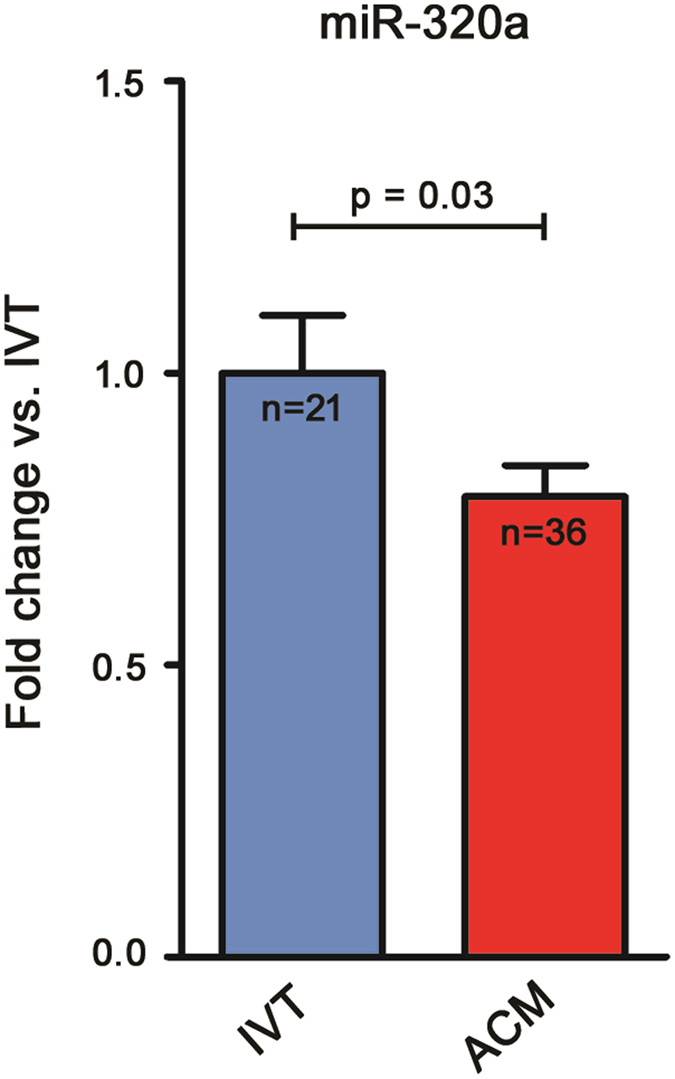
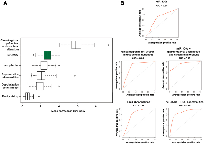
Diagnosis of Arrhythmogenic CardioMyopathy (ACM) is challenging and often late after disease onset. No circulating biomarkers are available to date. Given their involvement in several cardiovascular diseases, plasma microRNAs warranted investigation as potential non-invasive diagnostic tools in ACM. We sought to identify circulating microRNAs differentially expressed in ACM with respect to Healthy Controls (HC) and Idiopathic Ventricular Tachycardia patients (IVT), often in differential diagnosis. ACM and HC subjects were screened for plasmatic expression of 377 microRNAs and validation was performed in 36 ACM, 53 HC, 21 IVT. Variable importance in data partition was estimated through Random Forest analysis and accuracy by Receiver Operating Curves. Plasmatic miR-320a showed 0.53 ± 0.04 fold expression difference in ACM vs. HC (p < 0.01). A similar trend was observed when comparing ACM (n = 13) and HC (n = 17) with athletic lifestyle, a ACM precipitating factor. Importantly, ACM patients miR-320a showed 0.78 ± 0.05 fold expression change vs. IVT (p = 0.03). When compared to non-invasive ACM diagnostic parameters, miR-320a ranked highly in discriminating ACM vs. IVT and it increased their accuracy. Finally, miR-320a expression did not correlate with ACM severity. Our data suggest that miR-320a may be considered a novel potential biomarker of ACM, specifically useful in ACM vs. IVT differentiation.


Conflict of interest statement

The authors declare that they have no competing interests.

Figures

Figure 1
Figure 1
Non-invasive diagnostic tests in an Arrhythmogenic CardioMyopathy (ACM) patient. (A) Baseline 12-lead ECG shows regular sinus rhythm with inverted T waves in the precordial leads from V1 to V4. (B) Four-chamber echocardiographic imaging shows enlarged right ventricular diastolic volumes. Cardiac magnetic resonance: four-chamber short-axis (C) and long axis (D) images reveal dilated right ventricle with segmental kinesis abnormalities, mainly involving the inferolateral wall.
Figure 2
Figure 2
miR-320a shows a lower plasma expression in Arrhythmogenic CardioMyopathy (ACM) patients when compared to Healthy Controls (HC). Relative expression of plasma miR-320a in ACM patients (n = 36) with respect to HC (n = 53); the mean expression value in HC was arbitrarily set to one (HC 1.00 ± 0.11; ACM 0.53 ± 0.04; p < 0.01, t-test).
Figure 3
Figure 3
miR-320a plasmatic expression is not influenced by athletic lifestyle. Plasmatic relative expression of miR-320a in non-athlete Healthy Controls (HC; n = 36), athlete HC (n = 17), non-athlete Arrhythmogenic CardioMyopathy patients (ACM; n = 23), and athlete ACM patients (n = 13). Results are shown with respect to the mean expression of miR-320a in non-athlete HC, arbitrarily set to one. Kruskal-Wallis followed by Dunns test was used to determine that no significant difference is observed between non-athlete and athlete HC subgroups (1.00 ± 0.22 vs. 1.16 ± 0.13; p = 0.10) and between non-athlete and athlete ACM subgroups (0.57 ± 0.04 vs. 0.55 ± 0.07; p = 0.60). HC vs. ACM differences are confirmed both between the non-athlete (1.00 ± 0.22 vs. 0.57 ± 0.04; p = 0.05) and athlete (1.16 ± 0.13 vs. 0.55 ± 0.07; p < 0.01) subgroups.
Figure 4
Figure 4
miR-320a shows a lower plasmatic expression in Arrhythmogenic CardioMyopathy (ACM) patients when compared to patients affected by Idiopathic Ventricular Tachycardia (IVT). Plasma relative expression of miR-320a, in ACM patients (n = 36), with respect to mean expression in IVT patients (n = 21), arbitrarily set to one. (IVT 1 ± 0.10; ACM 0.78 ± 0.05; p = 0.03, Mann-Whitney).
Figure 5
Figure 5
Importance of non-invasive ACM established diagnostic variables and miR-320a in Arrhythmogenic CardioMyopathy (ACM) vs. Idiopathic Ventricular Tachycardia (IVT) classification. (A) Boxplots show the distribution of mean decreases in Gini index for non-invasive ACM diagnostic criteria and miR-320a (green boxplot), ranked based on the importance in data partition as calculated by the Random Forests procedure. Data are shown as median[Q1–Q3]: global/regional dysfunction and structural alteration, 5.75[5.40–6.39]; miR-320a, 2.72[2.43–3.12]; arrhythmias, 2.45[2.10–2.79]; repolarization abnormalities, 2.17[1.68–2.59]; depolarization abnormalities, 2.03[1.65–2.38]; family history, 0.66[0.48–0.86]. (B) The Area Under the Curves (AUC) of the ROC analysis was used to evaluate diagnostic accuracy of selected variables used to classify ACM vs. IVT patients and the added value by circulating miR-320a expression. ECG abnormalities: depolarization and repolarization abnormality criteria.

References

    1. Pilichou K, et al. Arrhythmogenic cardiomyopathy. Orphanet J Rare Dis. 2016;11:33. doi: 10.1186/s13023-016-0407-1. - DOI - PMC - PubMed
    1. Corrado D, et al. Trends in sudden cardiovascular death in young competitive athletes after implementation of a preparticipation screening program. JAMA. 2006;296:1593–1601. doi: 10.1001/jama.296.13.1593. - DOI - PubMed
    1. Lazzarini E, et al. The ARVD/C genetic variants database: 2014 update. Hum Mutat. 2015;36:403–410. doi: 10.1002/humu.22765. - DOI - PubMed
    1. Garcia-Gras E, et al. Suppression of canonical Wnt/beta-catenin signaling by nuclear plakoglobin recapitulates phenotype of arrhythmogenic right ventricular cardiomyopathy. J Clin Invest. 2006;116:2012–2021. doi: 10.1172/JCI27751. - DOI - PMC - PubMed
    1. Deb A. Cell-cell interaction in the heart via Wnt/beta-catenin pathway after cardiac injury. Cardiovasc Res. 2014;102:214–223. doi: 10.1093/cvr/cvu054. - DOI - PMC - PubMed

Publication types

MeSH terms