

LINE-1 hypomethylation is not a common event in preneoplastic stages of gastric carcinogenesis
- PMID: 28684753
- PMCID: PMC5500474
- DOI: 10.1038/s41598-017-05143-0
LINE-1 hypomethylation is not a common event in preneoplastic stages of gastric carcinogenesis
Abstract
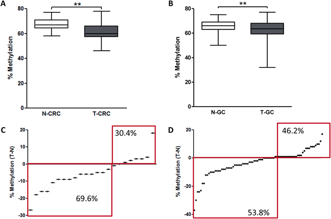
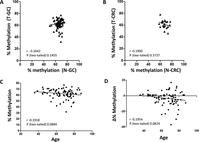
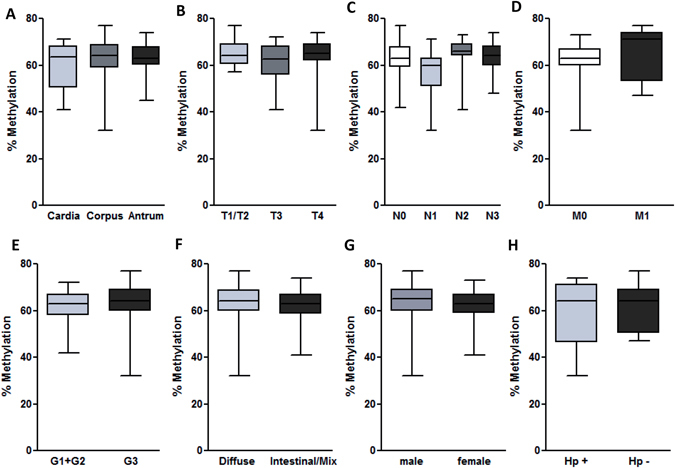
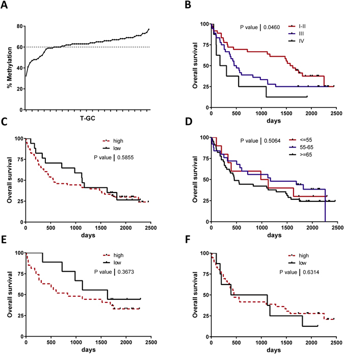
LINE-1 hypomethylation is widely accepted as marker for global genomic DNA hypomethylation, which is a frequent event in cancer. The aim of the study was to evaluate LINE-1 methylation status at different stages of gastric carcinogenesis and evaluate its prognostic potential in clinical settings. LINE-1 methylation was analyzed in 267 tissue samples by bisulfite pyrosequencing including primary colorectal cancer tissues (T-CRC) with corresponding adjacent colon mucosa (N-CRC), gastric cancer tissues (T-GC) with corresponding gastric mucosa (N-GC), normal gastric tissues (N), chronic non-atrophic and atrophic gastritis (CG). LINE-1 methylation level was lower in both T-GC and T-CRC when compared to paired adjacent tissues. No difference was observed for LINE-1 methylation status between patients with normal gastric mucosa, CG and N-GC. LINE-1 methylation in T-GC but not N-GC tended to correlate with age. Subgroup stratification analysis did not reveal significant differences in LINE-1 methylation status according to tumor stage, anatomical location, histological subtype, differentiation grade. We observed similar overall survival data between patients with high or low LINE-1 levels. In summary, LINE-1 hypomethylation is a characteristic feature in GC but not very common in early preneoplastic stages of gastric carcinogenesis. Prognostic role of LINE-1 hypomethylation in GC patients could not be confirmed in this cohort.
Conflict of interest statement
The authors declare that they have no competing interests.
Figures

Similar articles
-
Epigenetic silencing of miR-137 is a frequent event in gastric carcinogenesis.Mol Carcinog. 2016 Apr;55(4):376-86. doi: 10.1002/mc.22287. Epub 2015 Feb 7. Mol Carcinog. 2016. PMID: 25663388
-
Global DNA hypomethylation is an early event in Helicobacter pylori-related gastric carcinogenesis.J Clin Pathol. 2011 Aug;64(8):677-82. doi: 10.1136/jcp.2010.087858. Epub 2011 May 26. J Clin Pathol. 2011. PMID: 21617174
-
ALU and LINE-1 hypomethylations in multistep gastric carcinogenesis and their prognostic implications.Int J Cancer. 2012 Sep 15;131(6):1323-31. doi: 10.1002/ijc.27369. Epub 2012 Jan 3. Int J Cancer. 2012. PMID: 22120154
-
DNA methylation as a molecular biomarker in gastric cancer.Epigenomics. 2015;7(3):475-86. doi: 10.2217/epi.15.4. Epigenomics. 2015. PMID: 26077432 Review.
-
LINE-1 methylation level and prognosis in pancreas cancer: pyrosequencing technology and literature review.Surg Today. 2017 Dec;47(12):1450-1459. doi: 10.1007/s00595-017-1539-1. Epub 2017 May 23. Surg Today. 2017. PMID: 28536860 Review.
Cited by
-
Impact of LINE-1 hypomethylation on the clinicopathological and molecular features of colorectal cancer patients.PLoS One. 2018 May 24;13(5):e0197681. doi: 10.1371/journal.pone.0197681. eCollection 2018. PLoS One. 2018. PMID: 29795620 Free PMC article.
-
Standardization of DNA amount for bisulfite conversion for analyzing the methylation status of LINE-1 in lung cancer.PLoS One. 2021 Aug 17;16(8):e0256254. doi: 10.1371/journal.pone.0256254. eCollection 2021. PLoS One. 2021. PMID: 34403448 Free PMC article.
-
Tumor DNA hypomethylation of LINE-1 is associated with low tumor grade of breast cancer in Tunisian patients.Oncol Lett. 2020 Aug;20(2):1999-2006. doi: 10.3892/ol.2020.11745. Epub 2020 Jun 17. Oncol Lett. 2020. PMID: 32724446 Free PMC article.
-
Fusobacterium nucleatum is associated with worse prognosis in Lauren's diffuse type gastric cancer patients.Sci Rep. 2020 Oct 1;10(1):16240. doi: 10.1038/s41598-020-73448-8. Sci Rep. 2020. PMID: 33004953 Free PMC article.
-
Microbial composition of tumorous and adjacent gastric tissue is associated with prognosis of gastric cancer.Sci Rep. 2023 Mar 21;13(1):4640. doi: 10.1038/s41598-023-31740-3. Sci Rep. 2023. PMID: 36944721 Free PMC article.
References
-
- Jemal, A. et al. Global cancer statistics. CA. Cancer J. Clin. 61, 69–90. - PubMed
-
- Malfertheiner P, Link A, Selgrad M. Helicobacter pylori: perspectives and time trends. Nat. Rev. Gastroenterol. Hepatol. 2014;11:628–638. - PubMed
-
- Correa P. A human model of gastric carcinogenesis. Cancer Res. 1988;48:3554–60. - PubMed
Publication types
MeSH terms
LinkOut - more resources
Full Text Sources
Other Literature Sources
Medical
Miscellaneous
