. 2017 Jul 6;7(1):4793.
doi: 10.1038/s41598-017-04116-7.

Hepatocyte nuclear factor-1beta enhances the stemness of hepatocellular carcinoma cells through activation of the Notch pathway

Affiliations

Hepatocyte nuclear factor-1beta enhances the stemness of hepatocellular carcinoma cells through activation of the Notch pathway

Jing-Ni Zhu et al. Sci Rep. .

Abstract

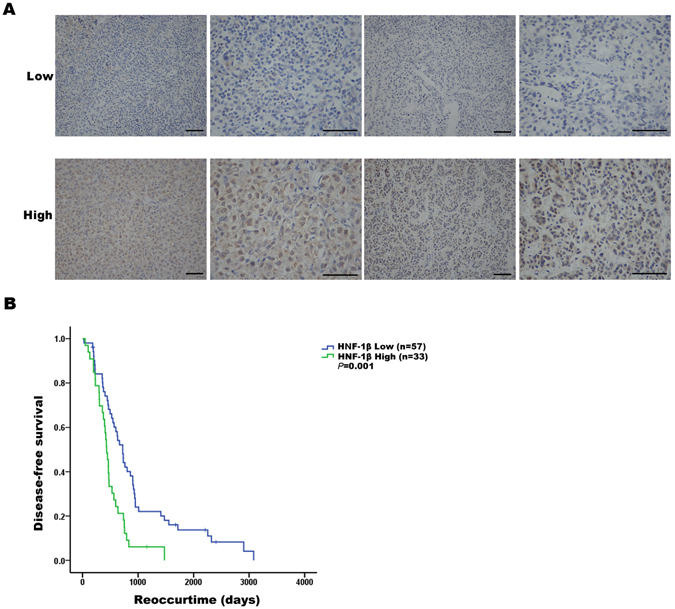
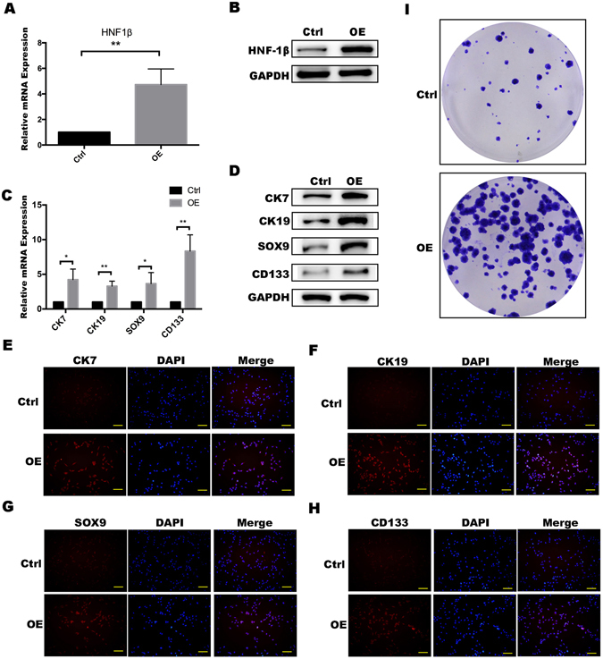
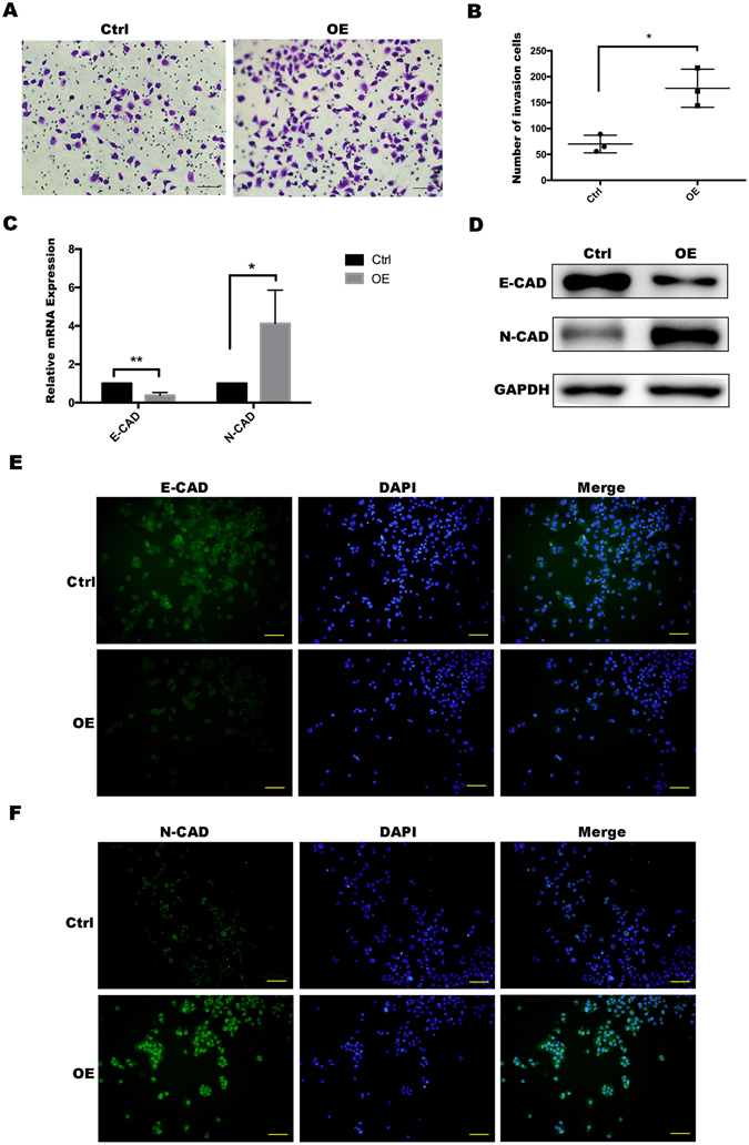
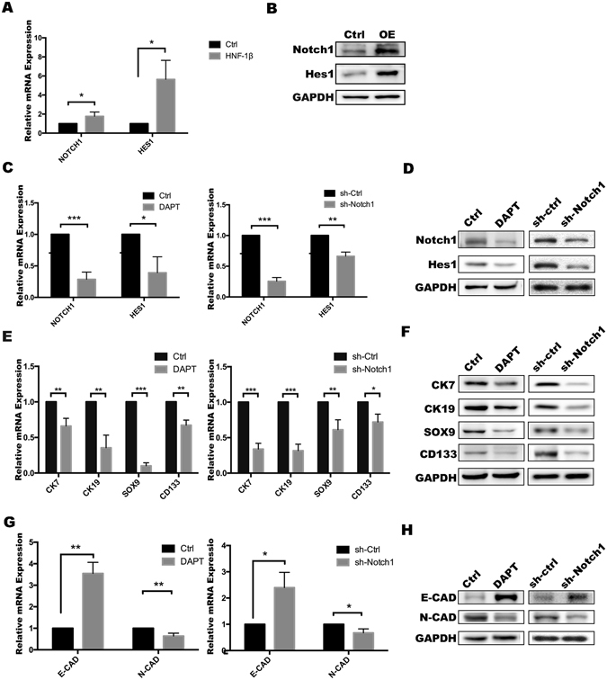
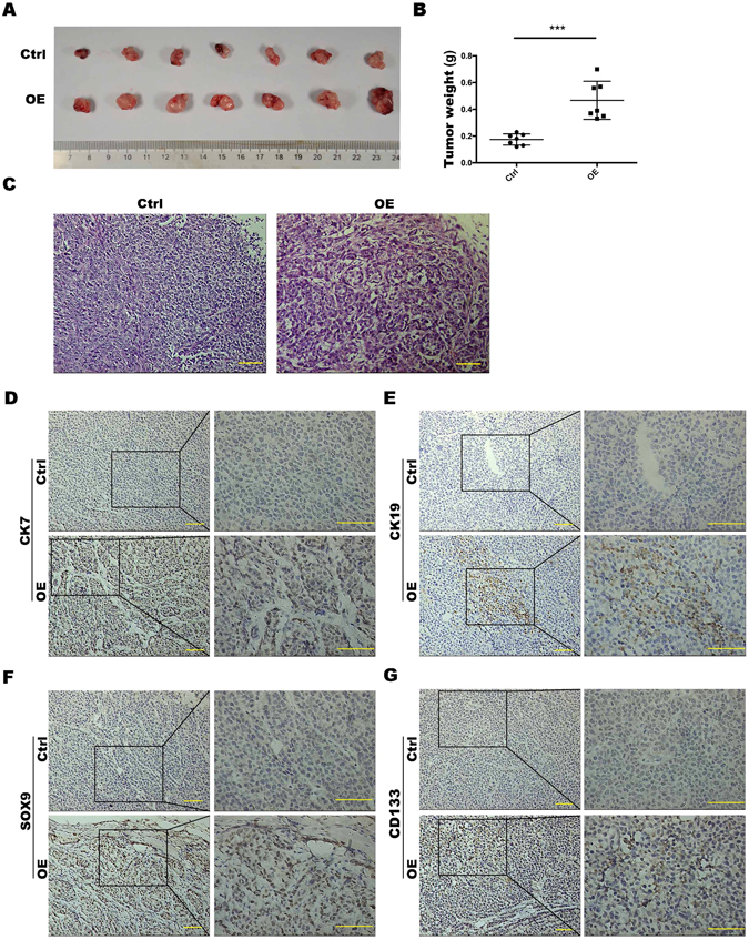
Hepatocyte nuclear factor-1beta plays an important role in the development and progression of liver cancer. In recent years, the expression of HNF-1β has been reported to be associated with risk for a variety of cancers. The purpose of this study is to investigate whether the expression of HNF-1β promotes the malignancy of HCC and its mechanism. We retrospectively investigated the expression of HNF-1β in 90 patients with hepatocellular carcinoma and found that the high expression of HNF-1β indicated poor prognosis. We overexpressed HNF-1β in liver cancer cell lines and found the expression of liver progenitor cell markers and stemness were upregulated. The invasion ability and epithelial-mesenchymal transition (EMT)-associated genes were also significantly higher in liver cancer cells overexpressing HNF-1β than in the control group. A mechanistic study suggested the activation of the Notch signalling pathway probably plays a key role downstream of HNF-1β. More importantly, HNF-1β promoted tumourigenesis of HCC cells in vivo. In conclusion, high expression of HNF-1β not only promoted the de-differentiation of HCC cells into liver cancer stem cells through activating the Notch pathway but also enhanced the invasive potential of HCC cells and EMT occurrence, which would contribute to the enhancement of cell migration and invasion.


Conflict of interest statement

The authors declare that they have no competing interests.

Figures

Figure 1
Figure 1
The expression of HNF-1β was negatively correlated with the prognosis of patients with HCC. (A) HNF-1β immunohistochemical staining patterns in HCC tissues. According to the HNF-1β expression level, HCC patients were divided into an HNF-1β low expression group (the top row) and high expression group (the bottom row). (scale bar = 100 μm). (B) Kaplan-Meier analysis of HNF-1β expression in HCC patients. Low expression of HNF-1β in HCC tissues was associated with prolonged disease-free survival. The P value calculated by the log-rank test is indicated (P = 0.001).
Figure 2
Figure 2
PLC/RF5 HCC cells with HNF-1β overexpression strongly expressed liver progenitor cell markers. (A,B) Detection of HNF-1β expression in HCC cells and HNF-1β-overexpressed HCC cells. OE: HNF-1β overexpression. (C,D) RT-PCR and western blotting were performed to detect the expression of the HPC markers (CK7, CK19, SOX9 and CD133). (*P < 0.05, **P < 0.01) Mean ± S.E.M. (E–H) Immunofluorescence staining was used to identify the expression level of the phenotypes (CK7, CK19, SOX9 and CD133) (scale bar = 100 μm). (I) A colony formation assay was used to detect the difference in the stemness between HCC cells and HNF-1β-overexpressed HCC cells.
Figure 3
Figure 3
HNF-1β overexpression promoted invasion of PLC/RF5 HCC cells. (A,B) Transwell invasion assay was used to observe the invasion ability of the HCCs and the HNF-1β-overexpressed HCC cells. (C,D) RT-PCR and western blotting was performed to detect the expression of the EMT markers (E-cadherin and N-cadherin) in HCC cells and the HNF-1β-overexpressed HCC cells. (*P < 0.05, **P < 0.01) Mean ± S.E.M. (E,F) Immunofluorescence staining was used to identify the expression level of the EMT markers (E-CAD and N-CAD) (scale bar = 100 μm).
Figure 4
Figure 4
HNF-1β upregulated expression of Notch signalling-related genes in PLC/RF5 HCC cells. (A,B) The activation of NOTCH1 and HES1 in HCC cells was detected by RT-PCR and western blotting analysis. (C,D) The inhibition of the Notch pathway by DAPT or shRNA was confirmed using RT-PCR and western blotting. (E,F) RT-PCR and western blotting were used to determine the expression of CK7, CK19, SOX9 and CD133 after treatment with DAPT or Notch1 shRNA in HNF-1β-overexpressed HCC cells. (G,H) The expression of EMT markers was detected by RT-PCR and western blotting. (*P < 0.05, **P < 0.01, *** P < 0.001) Mean ± S.E.M. OE: HNF-1β overexpression. sh-Ctrl: negative control shRNA. sh-Notch1: Notch1 shRNA.
Figure 5
Figure 5
HNF-1β overexpression enhanced the ability of HCC cells to form tumours in vivo. (A,B) Tumour size of nude mice injected with PLC/RF5 HCC cells or HNF-1β-overexpressed PLC/RF5 cells (P < 0.001, mean ± S.E.M). (C) Representative H&E images of tumours. (D–G) Representative IHC images of CK7, CK19, SOX9 and CD133 of tumours in the Ctrl group and HNF-1β-overexpressed group (scale bar = 100 μm). n = 7.

References

    1. Woo HG, et al. Identification of a cholangiocarcinoma-like gene expression trait in hepatocellular carcinoma. Cancer research. 2010;70:3034–3041. doi: 10.1158/0008-5472.CAN-09-2823. - DOI - PMC - PubMed
    1. Kim GJ, Kim H, Park YN. Increased expression of Yes-associated protein 1 in hepatocellular carcinoma with stemness and combined hepatocellular-cholangiocarcinoma. PloS one. 2013;8:e75449. doi: 10.1371/journal.pone.0075449. - DOI - PMC - PubMed
    1. Zhang M, Lee AV, Rosen JM. The Cellular Origin and Evolution of Breast Cancer. Cold Spring Harb Perspect Med. 2017 - PMC - PubMed
    1. Nolan KD, Kaur J, Isaacs JS. Secreted heat shock protein 90 promotes prostate cancer stem cell heterogeneity. Oncotarget. 2016 - PMC - PubMed
    1. Gupta PB, Chaffer CL, Weinberg RA. Cancer stem cells: mirage or reality? Nat Med. 2009;15:1010–1012. doi: 10.1038/nm0909-1010. - DOI - PubMed

Publication types

MeSH terms