Endovascular Approach to Glomus Jugulare Tumors
- PMID: 28685005
- PMCID: PMC5487373
- DOI: 10.12659/PJR.901141
Endovascular Approach to Glomus Jugulare Tumors
Abstract
Background: Paragangliomas are benign neuroendocrine tumors derived from the glomus cells of the vegetative nervous system. Typically, they are located in the region of the jugular bulb and middle ear. The optimal management is controversial and can include surgical excision, stereotactic radiosurgery and embolization.
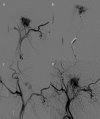
Case report: We report the endovascular approach to three patients harboring glomus jugulare paragangliomas. In all cases incomplete occlusion of the lesions was achieved and recanalization in the follow-up period was revealed. Two patients presented no clinical improvement and the remaining one experienced a transient withdrawal of tinnitus.
Conclusions: It is technically difficult to achieve complete obliteration of glomus jugulare tumors with the use of embolization and the subtotal occlusion poses a high risk of revascularization and is not beneficial in terms of alleviating clinical symptoms.
Keywords: Embolization, Therapeutic; Glomus Jugulare; Glomus Jugulare Tumor; Paraganglioma.
Figures


References
-
- Chen PG, Nguyen JH, Payne SC, et al. Treatment of glomus jugulare tumors with gamma knife radiosurgery. Laryngoscope. 2010;120:1856–62. - PubMed
-
- Genç A, Bicer A, Abacioglu U, et al. Gamma knife radiosurgery for the treatment of glomus jugulare tumors. J Neurooncol. 2010;97:101–8. - PubMed
-
- Winn HR, Youmans JR, editors. Youmans neurological surgery. 5th ed. Philadelphia: Saunders; 2004.
-
- Greenberg MS, Greenberg MS, editors. Handbook of neurosurgery. 7th ed. Tampa, FL: Greenberg Graphics; 2010.
Publication types
LinkOut - more resources
Full Text Sources
Other Literature Sources
