. 2017 Dec;4(1):19.
doi: 10.1186/s40658-017-0186-3. Epub 2017 Jul 6.

Design and implementation of a prototype head and neck phantom for the performance evaluation of gamma imaging systems

Affiliations

Design and implementation of a prototype head and neck phantom for the performance evaluation of gamma imaging systems

Mohammed S Alqahtani et al. EJNMMI Phys. 2017 Dec.

Abstract

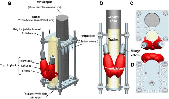
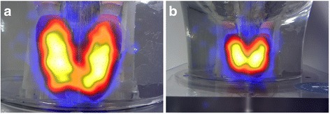
Background: A prototype anthropomorphic head and neck phantom has been designed to simulate the adult head and neck anatomy including some internal organs and tissues of interest, such as thyroid gland and sentinel lymph nodes (SLNs). The design of the head and neck phantom includes an inner jig holding the simulated SLNs and thyroid gland. The thyroid gland structure was manufactured using three-dimensional (3D) printing taking into consideration the morphology and shape of a healthy adult thyroid gland.

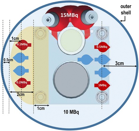
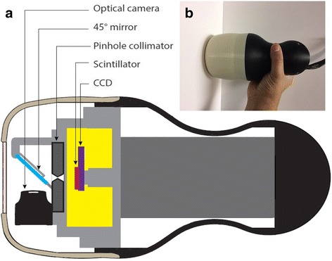
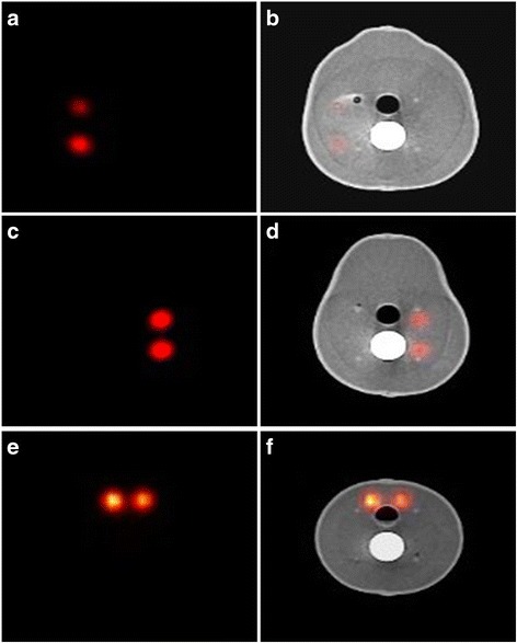
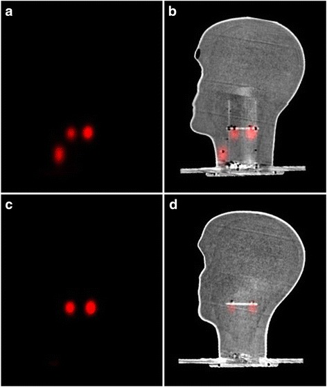
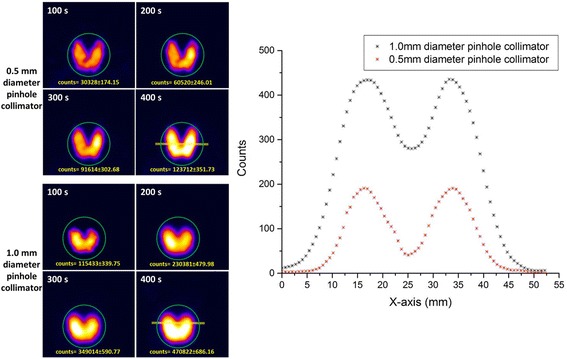
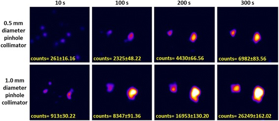
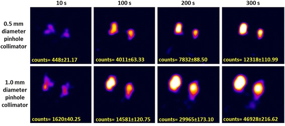
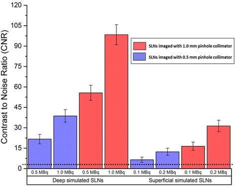
Result: The head and neck phantom was employed to simulate a situation where there are four SLNs distributed at two different vertical levels and at two depths within the neck. Contrast to noise ratio (CNR) calculations were performed for the detected SLNs at an 80 mm distance between both pinhole collimators (0.5 and 1.0 mm diameters) and the surface of the head and neck phantom with a 100 s acquisition time. The recorded CNR values for the simulated SLNs are higher when the hybrid gamma camera (HGC) was fitted with the 1.0 mm diameter pinhole collimator. For instance, the recorded CNR values for the superficially simulated SLN (15 mm depth) containing 0.1 MBq of 99mTc using 0.5 and 1.0 mm diameter pinhole collimators are 6.48 and 16.42, respectively (~87% difference). Gamma and hybrid optical images were acquired using the HGC for the simulated thyroid gland. The count profiles through the middle of the simulated thyroid gland images provided by both pinhole collimators were obtained. The HGC could clearly differentiate the individual peaks of both thyroid lobes in the gamma image produced by the 0.5-mm pinhole collimator. In contrast, the recorded count profile for the acquired image using the 1.0-mm-diameter pinhole collimator showed broader peaks for both lobes, reflecting the degradation of the spatial resolution with increasing the diameter of the pinhole collimator.

Conclusions: This anthropomorphic head and neck phantom provides a valuable tool for assessing the imaging ability of gamma cameras used for imaging the head and neck region. The standardisation of test phantoms for SFOV gamma systems will provide an opportunity to collect data across various medical centres. The phantom described is cost effective, reproducible, flexible and anatomically representative.

Keywords: 3D printing; Anthropomorphic phantom; Head and neck phantom; SFOV gamma camera; SPECT; Sentinel lymph nodes detection; Thyroid phantom; Thyroid scan.


Conflict of interest statement

Competing interests

The authors declare that they have no competing interests.

Publisher’s Note

Springer Nature remains neutral with regard to jurisdictional claims in published maps and institutional affiliations.

Figures

Fig. 1
Fig. 1
a Photograph of the head and neck phantom. b The internal jig with the attached thyroid phantom from anterolateral view b
Fig. 2
Fig. 2
Schematics of the internal jig showing the mounted simulated thyroid gland (red) and simulated sentinel lymph nodes (SLNs). The shape and position of the thyroid phantom relative to the simulated trachea: anterolateral (a), anterior (b), superior (c), and inferior (d) views
Fig. 3
Fig. 3
A schematic diagram showing a cross section through the neck region of the phantom; it shows the position, lateral depth and amount of radioactivity in the simulated lymph nodes
Fig. 4
Fig. 4
a A schematic diagram for the hybrid gamma camera (HGC) showing its internal structure. b Photograph of the HGC
Fig. 5
Fig. 5
Coronal SPECT and SPECT-CT images showing the position of the simulated SLNs (0.1–0.5 MBq) and the simulated thyroid gland (15 MBq) within the phantom (a, b); CT and SPECT-CT images of the midsagittal plane of the head and neck phantom showing the simulated thyroid gland, trachea and cervical spine (c, d)
Fig. 6
Fig. 6
SPECT and SPECT-CT images in the transverse plane representing the anatomical structure of the head and neck phantom; images a and b show two superficially low activity uptake simulated SLNs in gamma and hybrid modes (0.1 and 0.2 MBq, respectively). Images c and d show two deeper suited, higher activity uptake simulated SLNs in gamma and hybrid modes (0.5 and 1.0 MBq, respectively). Images e and f show the thyroid level SPECT and SPECT-CT images in the neck region (15 MBq)
Fig. 7
Fig. 7
SPECT and SPECT-CT images in the sagittal plane showing the deeper simulated SLNs (0.5–1.0 MBq) and the simulated thyroid gland in a and b; images c and d show the simulated superficial SLNs at the parotid gland level (0.1–0.2 MBq)
Fig. 8
Fig. 8
Left-hand side: planar HGC gamma images of the simulated thyroid gland at a 120 mm collimator-to-surface distance acquired with different acquisition times ranging from 100 to 400 s using both pinhole collimators (0.5 and 1.0 mm diameter). Right-hand side: count profiles plot for the data acquired from anterior gamma images for the simulated thyroid gland (400 s) using 0.5 and 1.0 mm diameter pinhole collimators. The yellow line in both thyroid gamma images (400 s) represents the cross-section area of the acquired data for both count profiles
Fig. 9
Fig. 9
Hybrid HGC images of the simulated thyroid gland at a distance of 100 mm (a) and 200 mm (b) from the phantom surface
Fig. 10
Fig. 10
Planar HGC gamma images for the two simulated, superficial SLNs (0.1 and 0.2 MBq) at an 80 mm collimator-to-surface distance utilising both pinhole collimators (0.5 and 1.0 mm diameter); the acquisition time varied between 10 and 300 s
Fig. 11
Fig. 11
Planar HGC gamma images for the two simulated, deep SLNs (0.5 and 1.0 MBq) at an 80 mm collimator-to-surface distance; both pinhole collimators (0.5 and 1.0 mm diameter) were used, and the acquisition time varied between 10 and 300 s
Fig. 12
Fig. 12
Bar chart showing the recorded contrast to noise ratio (CNR) values of different radioactivity concentrations for the simulated superficial and deep SLNs (i.e. 15 and 30 mm depth) for a 100 s acquisition time. Error bars represent the standard deviation of the CNR calculation for each node. The dotted line at CNR = 3 represents a threshold value of the Rose criterion of detectability

References

    1. Chen SL, Iddings DM, Scheri RP, Bilchik AJ. Lymphatic mapping and sentinel node analysis: current concepts and applications. CA Cancer J Clin. 2006;56:292–309. doi: 10.3322/canjclin.56.5.292. - DOI - PubMed
    1. Stoeckli SJ, Steinert H, Pfaltz M, Schmid S. Sentinel lymph node evaluation in squamous cell carcinoma of the head and neck. Otolaryngol Head Neck Surg. 2001;125:221–6. doi: 10.1067/mhn.2001.118074. - DOI - PubMed
    1. Hellingman D, de Wit-van der Veen LJ, Klop WM, Olmos RA. Detecting near-the-injection-site sentinel nodes in head and neck melanomas with a high-resolution portable gamma camera. Clin Nucl Med. 2015;40:e11–6. doi: 10.1097/RLU.0000000000000370. - DOI - PubMed
    1. Gershenwald JE, Thompson W, Mansfield PF, Lee JE, Colome MI, Tseng CH, et al. Multi-institutional melanoma lymphatic mapping experience: the prognostic value of sentinel lymph node status in 612 stage I or II melanoma patients. J Clin Oncol. 1999;17(3):976–83. doi: 10.1200/JCO.1999.17.3.976. - DOI - PubMed
    1. Lin D, Franc BL, Kashani-Sabet M, Singer MI. Lymphatic drainage patterns of head and neck cutaneous melanoma observed on lymphoscintigraphy and sentinel lymph node biopsy. Head Neck. 2006;28:249–55. doi: 10.1002/hed.20328. - DOI - PubMed

LinkOut - more resources