. 2017 Aug;27(8):1046-1064.
doi: 10.1038/cr.2017.88. Epub 2017 Jul 7.

Existing drugs as broad-spectrum and potent inhibitors for Zika virus by targeting NS2B-NS3 interaction

Affiliations

Existing drugs as broad-spectrum and potent inhibitors for Zika virus by targeting NS2B-NS3 interaction

Zhong Li et al. Cell Res. 2017 Aug.

Abstract

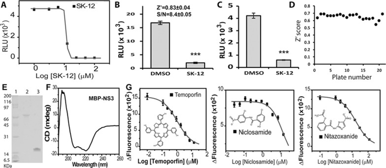
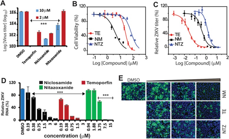
Recent outbreaks of Zika virus (ZIKV) highlight an urgent need for therapeutics. The protease complex NS2B-NS3 plays essential roles during flaviviral polyprotein processing, and thus represents an attractive drug target. Here, we developed a split luciferase complementation-based high-throughput screening assay to identify orthosteric inhibitors that directly target flavivirus NS2B-NS3 interactions. By screening a total of 2 816 approved and investigational drugs, we identified three potent candidates, temoporfin, niclosamide, and nitazoxanide, as flavivirus NS2B-NS3 interaction inhibitors with nanomolar potencies. Significantly, the most potent compound, temoporfin, not only inhibited ZIKV replication in human placental and neural progenitor cells, but also prevented ZIKV-induced viremia and mortality in mouse models. Structural docking suggests that temoporfin potentially binds NS3 pockets that hold critical NS2B residues, thus inhibiting flaviviral polyprotein processing in a non-competitive manner. As these drugs have already been approved for clinical use in other indications either in the USA or other countries, they represent promising and easily developed therapies for the management of infections by ZIKV and other flaviviruses.


Figures

Figure 1
Figure 1
The DENV2 NS2B/NS3 SLC assays. (A) The DENV2 NS2B/NS3 SLC constructs. NLuc aa 1-416-NS2B (named as NLuc-NS2B), NS2B-NLuc aa 1-416 (NS2B-NLuc), NLuc416-NS2B aa 49-66 (NLuc-E66stop), NS2B (aa 49-66)-NLuc416 (E66stop-NLuc), GST-NS3-CLuc aa 398-550 (GNC), and GST-CLuc (aa 398-550)-NS3 (GCN). (B) The positions of NLuc and CLuc are important. Equal concentrations (100 nM) of each pair of NS2B/NS3 constructs were mixed (or alone) and incubated with luciferin substrate. ***P < 0.001. In all bar graphs, means and SD from triplicate experimental data were shown, unless otherwise specified. (C) Effects of detergent at 0.05% concentration. **P < 0.01; ***P < 0.001. (D) Dose-response of NLuc-E66stop/GCN pair. Nluc-E66stop at 20 nM was included in each experiment. Concentration of GCN was varied as indicated. ***P < 0.001. (E) The DENV2 MBP-NS3 fusion protein specifically inhibited the SLC by NLuc-E66stop and GCN (80 nM each). MBP-NS3 or MBP were at 3.25 μM each. ***P < 0.001. (F) Dose-response inhibition of the SLC signals from NLuc-E66stop and GCN by “cold” MBP-NS3. Experimental data were fitted using the sigmoidal function with the Origin6.0 software. (G) NS2B mutations greatly reduced SLC. GCN was paired with equal molar of NLuc-E66stop or NLuc-E66stop mutants (L51A, L53A, and V59A). ***P < 0.001.
Figure 2
Figure 2
HTS assay identified potent orthosteric protease inhibitors. (A) Dose-response inhibition of the SLC signals from NLuc-E66stop and GCN by SK-12. Experimental data were fitted using the sigmoidal function with the Origin6.0 software. (B) HTS parameters using purified NLuc-E66stop and GCN. NLuc-E66stop and GCN at 100 nM were used with DMSO or SK-12 (40 μM). n = 8. ***P < 0.001. (C) NS3 pockets were accessible to small molecule inhibitor when NS2B and NS3 were co-expressed. Plasmids of NLuc-E66stop and GCN were co-transformed into Escherichia coli BL21 (DE3). Cells were grown to OD600 of 0.6 and were induced by IPTG. Cells were continuously grown for 2 h, collected and resuspended in luciferase assay buffer. About 100 μl of cells was dispensed into a 96-well plate, incubated with 1% DMSO or SK-12 (40 μM) for 2 h, then mixed with substrate luciferin n = 8. ***P < 0.001. (D) Summary of HT screening of the NCGC Pharmaceutical Collection in all plates. Statistics were generated by averaging those of 20 plates that were calculated from 32 wells in each plate. (E) SDS-PAGE analysis of purified His-MBP-NS3 (lane 2) and His-NS2B (Lane 3). Lane 1, Bio-Rad broad range molecular weight (MW) standard. (F) CD spectrum of purified His-MBP-NS3. (G) Sigmoidal curve fittings of dose-response inhibitions of the His-NS2B/His-MBP-NS3 protease activities by drugs. Inset: schematic representations of identified drugs.
Figure 3
Figure 3
Drugs are potent inhibitors of ZIKV and DENV2. (A) Inhibition of DENV2 in viral reduction assay in A549 cells by temoporfin (TE), niclosamide (NM), and nitazoxanide (NTZ) at 10 and 2 μM concentrations. ***P < 0.001. (B) Cell viability assay. A549 cells were incubated with various concentrations of drugs and then assayed for viability at 48 h post incubation. Experimental data were fitted using a sigmoidal function. (C) Sigmoidal fittings of dose-dependent inhibition of ZIKV by drugs in A549 cells. Viral plaque reduction assay was used. (D) qRT-PCR analysis of inhibition of viral RNA from ZIKV-infected A549 cells by drugs. ***P < 0.001. (E) Immunofluorescence assay of inhibition of viral protein expression by drugs, using pan-flavivirus anti-E 4G2 antibody (green) (ATCC). Concentration for drugs: (1) niclosamide: 0.19, 0.57, and 1.67 μM; (2) temoporfin: 0.06, 0.56, and 5 μM; (3) nitazoxanide: 0.06, 0.56, and 5 μM; Nuclei (blue) were stained in all immunofluorescence assays (IFA) by the Hoechst stain solution.
Figure 4
Figure 4
Inhibition of ZIKV in cells relevant to ZIKV. (A, D) Viral plaque reduction assay for ZIKV-infected HPECs (A) and iPSC-derived hNPCs (D) by drugs. *P < 0.05; **P < 0.01; ***P < 0.001. (B, E) Immunofluorescence assays (IFA) of inhibition of viral protein expression for ZIKV-infected HPECs (B) and hNPCs (E) by drugs. For HPECs, temoporfin (0.06 μM), niclosamide (0.19 μM), and nitazoxanide (10 μM). For hNPCs, temoporfin (1.0 μM), niclosamide (0.83 μM), and nitazoxanide (3.3 μM). (C, F) qRT-PCR analysis of inhibition of viral RNA of ZIKV-infected HPECs (C) and hNPCs (F) by drugs. ***P < 0.001. (G) qRT-PCR analysis of inhibition of viral RNA of ZIKV-infected iPSC HDF9 by drugs. ***P < 0.001. (H) Time-of-addition study of ZIKV inhibition in A549 cells by temoporfin (90 nM) and niclosamide (0.75 μM). ***P < 0.001.
Figure 5
Figure 5
In vivo antiviral activity of temoporfin against ZIKV. (A) Viremia was detected by RT-qPCR on day 2 post-ZIKV infection in 3-week-old Balb/C mice. Difference between temoporfin (n = 8) or vehicle (n = 7) treatment was analyzed by using the unpaired, two-tailed t-test. (B) Survival percentage for 4-week-old A129 mice infected with ZIKV and treated with temoporfin (n = 12) or vehicle (n = 10). Survival curves were compared using the Log-rank test.
Figure 6
Figure 6
Drugs directly bind to the NS3 protease domain and disrupt interactions between NS2B and NS3. (A) GST pull-down assay. GST-NS3 or the GST-tag (10 μg) was immobilized on the Glutathione sepharose-4B affinity beads (GE HealthCare). The FLAG-tagged NS2B (10 μg) was incubated with the beads for 2 h, and subjected to western blots (WB), using anti-FLAG (Genscript) and anti-GST antibodies (GE HealthCare). (B) Dose-dependent inhibition of NS2B-NS3 interactions by drugs, using the GST pull-down assay. The assay was performed the same as in A, except that two-fold dilution series of drugs were incubated with the GST-NS3 beads overnight prior to incubation with the FLAG-NS2B. Bottom panels showed normalized binding of FLAG-tag NS2B to GST-NS3. The binding of NS2B to NS3 in the absence of each drug (DMSO control) was set as 100%. The relative binding of NS2B to NS3 in the presence of each drug was normalized to the DMSO control. n = 3. (C) PTSA for binding of drugs to the MBP-NS3 protein. ΔTm was defined as Tm−drugTmDMSO. (D) SPR sensorgrams of kinetic data for the binding of drugs to refolded NS3. The refolded His-NS3 was coupled to a ProteOn GLH sensor chip (∼15 000 RU). Each drug with three-fold dilutions was injected. Global fitting of data to a 1:1 binding model is shown in dark black.
Figure 7
Figure 7
Drugs inhibit viral polyprotein precursor (PP) processing. (A) Lineweaver-Burk plot of kinetics experimental data for inhibition of the His-NS2B/His-MBP-NS3 protease complex by drugs. The DENV2 MBP-NS3 (100 nM) was mixed with temoporfin (3, 1.5, and 0.75 μM), niclosamide (30, 15, and 7.5 μM), or nitazoxanide (30, 15, and 7.5 μM) for 30 min. The DENV2 His-NS2B (1 μM) was added together with the Abz substrate at various concentrations (800-25 μM in two-fold dilutions). (B-D) Western blots (WB) analysis of dose-dependent inhibition of ZIKV NS3 expression by temoporfin (B), niclosamide (C), and nitazoxanide (D) using the GTX133309 ZIKV α-NS3 antibody (GeneTex) (left panel), respectively. The experiment was performed at the 48 h time point. Middle panel, NS3 expression (lower bands) normalized to the GAPDH loading control. Right panel, accumulated PP normalized to the DMSO control. **P < 0.01; ***P < 0.001. (E) MS/MS spectra obtained from the fragmentation of the precursor ion at m/z corresponding to representative ZIKV peptides. Fragment ions corresponding to y- and b-ions were observed (red lines).
Figure 8
Figure 8
Induced fit docking of drugs to the NS2B 2B51 and 2B53 pockets on NS3pro. (A, B) Ribbon presentation of temoporfin (green) docked into NS3pro of DENV3 (PDB: 3U1I) (A) and ZIKV (PDB: 5LC0) (B), respectively. NS3pro β-strand hairpin loops with residues 25-36 (DENV3) or 1 025-1 036 (ZIKV) are shown in orange and the loops with residues 56-67 (DENV3) or 1 056-1 067 (ZIKV) are shown in yellow. Key interaction residues are highlighted in stick presentation. Hydrogen bonds are shown in purple dotted lines and π-π stacking is shown in blue dotted line.

References

    1. Calvet G, Aguiar RS, Melo AS, et al. Detection and sequencing of Zika virus from amniotic fluid of fetuses with microcephaly in Brazil: a case study. Lancet Infect Dis 2016; 16:653–660. - PubMed
    1. Chen LH, Hamer DH. Zika Virus: rapid spread in the western hemisphere. Ann Intern Med 2016; 164:613–615. - PubMed
    1. Govero J, Esakky P, Scheaffer SM, et al. Zika virus infection damages the testes in mice. Nature 2016; 540:438–442. - PMC - PubMed
    1. Ma W, Li S, Ma S, et al. Zika virus causes testis damage and leads to male infertility in mice. Cell 2016; 167:1511–1524. e15. - PubMed
    1. Miner JJ, Sene A, Richner JM, et al. Zika virus infection in mice causes panuveitis with shedding of virus in tears. Cell Rep 2016; 16:3208–3218. - PMC - PubMed

MeSH terms