

Simultaneous targeting of ATM and Mcl-1 increases cisplatin sensitivity of cisplatin-resistant non-small cell lung cancer
- PMID: 28686074
- PMCID: PMC5653185
- DOI: 10.1080/15384047.2017.1345391
Simultaneous targeting of ATM and Mcl-1 increases cisplatin sensitivity of cisplatin-resistant non-small cell lung cancer
Erratum in
-
Correction.Cancer Biol Ther. 2020 May 3;21(5):476. doi: 10.1080/15384047.2020.1733804. Epub 2020 Feb 24. Cancer Biol Ther. 2020. PMID: 32093521 Free PMC article. No abstract available.
-
Correction.Cancer Biol Ther. 2021 Jun 3;22(5-6):415. doi: 10.1080/15384047.2021.1937449. Epub 2021 Jul 12. Cancer Biol Ther. 2021. PMID: 34251975 Free PMC article. No abstract available.
Abstract
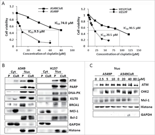
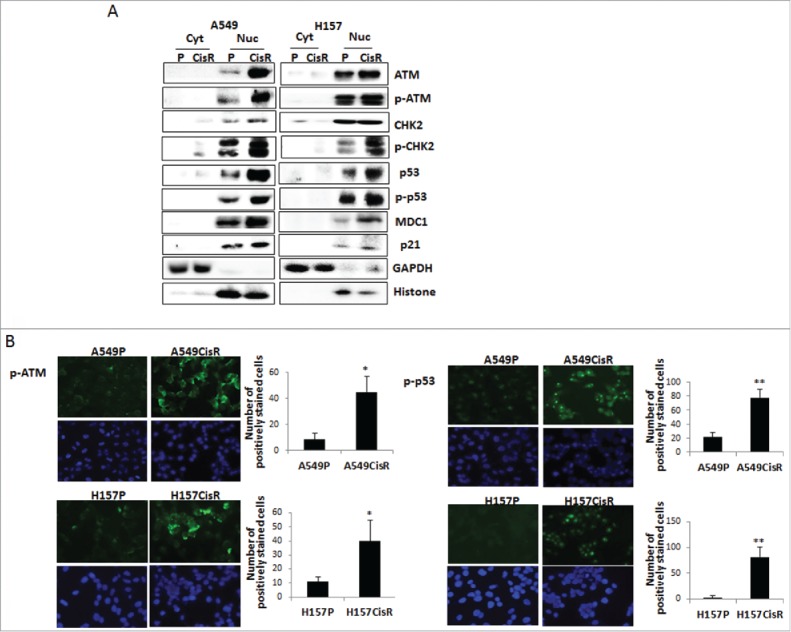
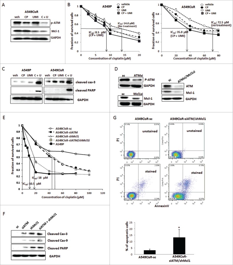
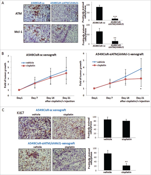
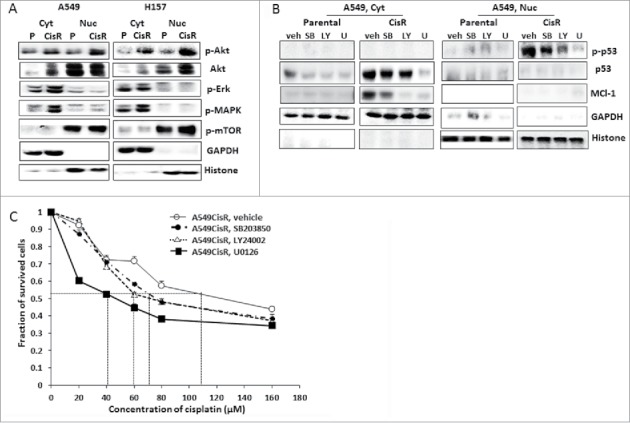
Development of cisplatin-resistance is an obstacle in non-small cell lung cancer (NSCLC) therapeutics. To investigate which molecules are associated with cisplatin-resistance, we analyzed expression profiles of several DNA repair and anti-apoptosis associated molecules in parental (A549P and H157P) and cisplatin-resistant (A549CisR and H157CisR) NSCLC cells. We detected constitutively upregulated nuclear ATM and cytosolic Mcl-1 molcules in cisplatin-resistant cells compared with parental cells. Increased levels of phosphorylated ATM (p-ATM) and its downstream molecules, CHK2, p-CHK2, p-53, and p-p53 were also detected in cisplatin-resistant cells, suggesting an activation of ATM signaling in these cells. Upon inhibition of ATM and Mcl-1 expression/activity using specific inhibitors of ATM and/or Mcl-1, we found significantly enhanced cisplatin-cytotoxicity and increased apoptosis of A549CisR cells after cisplatin treatment. Several A549CisR-derived cell lines, including ATM knocked down (A549CisR-siATM), Mcl-1 knocked down (A549CisR-shMcl1), ATM/Mcl-1 double knocked down (A549CisR-siATM/shMcl1) as well as scramble control (A549CisR-sc), were then developed. Higher cisplatin-cytotoxicity and increased apoptosis were observed in A549CisR-siATM, A549CisR-shMcl1, and A549CisR-siATM/shMcl1 cells compared with A549CisR-sc cells, and the most significant effect was shown in A549CisR-siATM/shMcl1 cells. In in vivo mice studies using subcutaneous xenograft mouse models developed with A549CisR-sc and A549CisR-siATM/shMcl1 cells, significant tumor regression in A549CisR-siATM/shMcl1 cells-derived xenografts was observed after cisplatin injection, but not in A549CisR-sc cells-derived xenografts. Finally, inhibitor studies revealed activation of Erk signaling pathway was most important in upregulation of ATM and Mcl-1 molcules in cisplatin-resistant cells. These studies suggest that simultaneous blocking of ATM/Mcl-1 molcules or downstream Erk signaling may recover the cisplatin-resistance of lung cancer.
Keywords: ATM; Mcl-1; cisplatin-resistance; cisplatin-sensitivity; non-small cell lung cancer.
Figures
Similar articles
-
Inhibition of ATM reverses EMT and decreases metastatic potential of cisplatin-resistant lung cancer cells through JAK/STAT3/PD-L1 pathway.J Exp Clin Cancer Res. 2019 Apr 8;38(1):149. doi: 10.1186/s13046-019-1161-8. J Exp Clin Cancer Res. 2019. PMID: 30961670 Free PMC article.
-
A FASN-TGF-β1-FASN regulatory loop contributes to high EMT/metastatic potential of cisplatin-resistant non-small cell lung cancer.Oncotarget. 2016 Aug 23;7(34):55543-55554. doi: 10.18632/oncotarget.10837. Oncotarget. 2016. PMID: 27765901 Free PMC article.
-
IL-6 signaling contributes to cisplatin resistance in non-small cell lung cancer via the up-regulation of anti-apoptotic and DNA repair associated molecules.Oncotarget. 2015 Sep 29;6(29):27651-60. doi: 10.18632/oncotarget.4753. Oncotarget. 2015. PMID: 26313152 Free PMC article.
-
Molecular Mechanisms of Chemoresistance Induced by Cisplatin in NSCLC Cancer Therapy.Int J Mol Sci. 2021 Aug 18;22(16):8885. doi: 10.3390/ijms22168885. Int J Mol Sci. 2021. PMID: 34445588 Free PMC article. Review.
-
Platinum-based targeted chemotherapies and reversal of cisplatin resistance in non-small cell lung cancer (NSCLC).Mutat Res. 2024 Jan-Jun;828:111856. doi: 10.1016/j.mrfmmm.2024.111856. Epub 2024 Mar 19. Mutat Res. 2024. PMID: 38520879 Review.
Cited by
-
Inhibition of ATM reverses EMT and decreases metastatic potential of cisplatin-resistant lung cancer cells through JAK/STAT3/PD-L1 pathway.J Exp Clin Cancer Res. 2019 Apr 8;38(1):149. doi: 10.1186/s13046-019-1161-8. J Exp Clin Cancer Res. 2019. PMID: 30961670 Free PMC article.
-
A novel necroptosis-related LncRNA signature for prediction of prognosis and therapeutic responses of head and neck squamous cell carcinoma.Front Pharmacol. 2022 Aug 9;13:963072. doi: 10.3389/fphar.2022.963072. eCollection 2022. Front Pharmacol. 2022. PMID: 36016575 Free PMC article.
-
DNA repair pathways and their roles in drug resistance for lung adenocarcinoma.Mol Biol Rep. 2021 Apr;48(4):3813-3825. doi: 10.1007/s11033-021-06314-z. Epub 2021 Apr 15. Mol Biol Rep. 2021. PMID: 33856604 Review.
-
Chemosensitizing activity of peptide from Lentinus squarrosulus (Mont.) on cisplatin-induced apoptosis in human lung cancer cells.Sci Rep. 2021 Feb 18;11(1):4060. doi: 10.1038/s41598-021-83606-1. Sci Rep. 2021. PMID: 33603033 Free PMC article.
-
Breathing New Life into the Mechanisms of Platinum Resistance in Lung Adenocarcinoma.Front Cell Dev Biol. 2020 May 8;8:305. doi: 10.3389/fcell.2020.00305. eCollection 2020. Front Cell Dev Biol. 2020. PMID: 32457904 Free PMC article. No abstract available.
References
-
- Cersosimo RJ. Lung cancer: a review. Am J Health-System Pharmacy: AJHP: Official J Am Society Health-System Pharmacists 2002; 59(7):611-42. Epub 2002/04/12; PMID:11944603 - PubMed
-
- Parsons A, Daley A, Begh R, Aveyard P. Influence of smoking cessation after diagnosis of early stage lung cancer on prognosis: systematic review of observational studies with meta-analysis. BMJ 2010; 340:b5569. Epub 2010/01/23; PMID:20093278; https://doi.org/10.1136/bmj.b5569 - DOI - PMC - PubMed
-
- Chang A. Chemotherapy, chemoresistance and the changing treatment landscape for NSCLC. Lung Cancer 2011; 71(1):3-10; PMID:20951465; https://doi.org/10.1016/j.lungcan.2010.08.022 - DOI - PubMed
-
- Galluzzi L, Vitale I, Michels J, Brenner C, Szabadkai G, Harel-Bellan A, Castedo M, Kroemer G. Systems biology of cisplatin resistance: past, present and future. Cell Death Disease 2014; 5:e1257; PMID:24874729; https://doi.org/10.1038/cddis.2013.428 - DOI - PMC - PubMed
-
- Galluzzi L, Senovilla L, Vitale I, Michels J, Martins I, Kepp O, Castedo M, Kroemer G. Molecular mechanisms of cisplatin resistance. Oncogene 2012; 31(15):1869-83; PMID:21892204; https://doi.org/10.1038/onc.2011.384 - DOI - PubMed
MeSH terms
Substances
LinkOut - more resources
Full Text Sources
Other Literature Sources
Medical
Research Materials
Miscellaneous