

Correlation between the position of hyoid bone and subregions of the pharyngeal airway space in lateral cephalometry and cone beam computed tomography
- PMID: 28686091
- PMCID: PMC8357209
- DOI: 10.2319/022217-133.1
Correlation between the position of hyoid bone and subregions of the pharyngeal airway space in lateral cephalometry and cone beam computed tomography
Abstract
Objective: To correlate the pharyngeal airway subregions with the positioning of the hyoid bone.
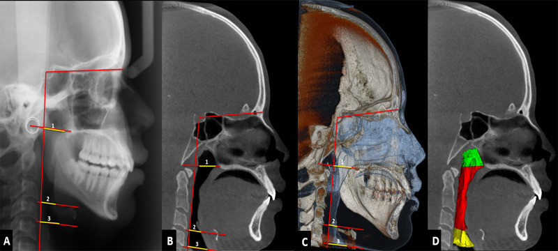
Material and methods: The study examined 107 lateral cephalometric (LC) and cone beam computed tomography (CBCT) images. Linear and volumetric measurements of the pharyngeal subregions were made and correlated to linear measurements using hyoid triangle analysis on images of LC and multiplanar (MPR) and three-dimensional (3D) reconstructions of CBCT.
Results: There was significant correlation between linear measurements of the pharyngeal subregions and hyoid bone position in LC images and in MPR and 3D reconstructions of the CBCT. Correlations were more frequent in the oropharynx and hypopharynx, especially for LC images. No correlations were observed between LC images or CBCT reconstructions and the volumetric measurements of the pharyngeal subregions and the position of the hyoid bone.
Conclusion: The hyoid bone position showed more correlations with oropharynx and hypopharynx airway measurements. The hyoid triangle method was not applicable to 3D images, since it showed a smaller number of measures correlated to the hyoid bone position.
Keywords: Cone beam computed tomography; Hyoid bone; Pharynx; Radiography.
Figures


Similar articles
-
Gender-related difference in the upper airway dimensions and hyoid bone position in Chinese Han children and adolescents aged 6-18 years using cone beam computed tomography.Acta Odontol Scand. 2015 Jul;73(5):391-400. doi: 10.3109/00016357.2014.978366. Epub 2015 Jan 28. Acta Odontol Scand. 2015. PMID: 25630980
-
Comparison of pharyngeal airway changes on plain radiography and cone-beam computed tomography after orthognathic surgery.J Oral Maxillofac Surg. 2011 Nov;69(11):e385-94. doi: 10.1016/j.joms.2011.03.015. Epub 2011 Jul 20. J Oral Maxillofac Surg. 2011. PMID: 21778015
-
Cone-beam computed tomography airway measurements: Can we trust them?Am J Orthod Dentofacial Orthop. 2019 Jul;156(1):53-60. doi: 10.1016/j.ajodo.2018.07.024. Am J Orthod Dentofacial Orthop. 2019. PMID: 31256838
-
Are three-dimensional airway evaluations obtained through computed and cone-beam computed tomography scans predictable from lateral cephalograms? A systematic review of evidence.Angle Orthod. 2017 Jan;87(1):159-167. doi: 10.2319/032516-243.1. Epub 2016 Jul 27. Angle Orthod. 2017. PMID: 27463700 Free PMC article.
-
Extraoral anatomy in CBCT – a literature review. Part 4: Pharyngocervical region.Swiss Dent J. 2020 Oct 12;130(10):768-784. doi: 10.61872/sdj-2020-10-01. Swiss Dent J. 2020. PMID: 33021766 Review.
Cited by
-
Three-dimensional pharyngeal airway and hyoid bone changes in skeletal class I malocclusion treated with extraction and non-extraction protocols: a comparative study of fixed orthodontic appliance and clear aligners.Clin Oral Investig. 2025 May 15;29(6):299. doi: 10.1007/s00784-025-06367-5. Clin Oral Investig. 2025. PMID: 40372546
-
Comparison of invisalign mandibular advancement and twin-block on upper airway and hyoid bone position improvements for skeletal class II children: a retrospective study.BMC Oral Health. 2023 Sep 13;23(1):661. doi: 10.1186/s12903-023-03295-2. BMC Oral Health. 2023. PMID: 37705022 Free PMC article.
-
Automated 3D segmentation of the hyoid bone in CBCT using nnU-Net v2: a retrospective study on model performance and potential clinical utility.BMC Med Imaging. 2025 Jul 1;25(1):217. doi: 10.1186/s12880-025-01797-9. BMC Med Imaging. 2025. PMID: 40596942 Free PMC article.
-
Cephalometric Evaluation in Patients with Obstructive Sleep Apnea undergoing Lateral Pharyngoplasty.Int Arch Otorhinolaryngol. 2024 Mar 6;28(2):e278-e287. doi: 10.1055/s-0043-1776718. eCollection 2024 Apr. Int Arch Otorhinolaryngol. 2024. PMID: 38618602 Free PMC article.
-
Study of Upper Pharyngeal Airway Dimension in Young Adults Visiting Orthodontic Department of a Dental College: A Descriptive Cross-sectional Study.JNMA J Nepal Med Assoc. 2021 Mar 31;59(235):271-275. doi: 10.31729/jnma.6293. JNMA J Nepal Med Assoc. 2021. PMID: 34506436 Free PMC article.
References
-
- Sheng CM, Lin LH, Su Y, Tsai HH. Developmental changes in pharyngeal airway depth and hyoid bone position from childhood to young adulthood. Angle Orthod. 2009;79:484–490. - PubMed
-
- Jiang YY. Correlation between hyoid bone position and airway dimensions in Chinese adolescents by cone beam computed tomography analysis. Int J Oral Maxillofac Surg. 2016;45:914–921. - PubMed
-
- McNamara JA. Influence of respiratory pattern on craniofacial growth. Angle Orthod. 1981;51:269–300. - PubMed
-
- Lenza MG, Lenza MM, Dalstra M, Melsen B, Cattaneo PM. An analysis of different approaches to the assessment of upper airway morphology: a CBCT study. Orthod Craniofac Res. 2010;13:96–105. - PubMed
MeSH terms
LinkOut - more resources
Full Text Sources
Other Literature Sources
