. 2018 Feb;16(2):200-208.
doi: 10.2450/2017.0176-16. Epub 2017 May 30.

Enhanced opsonisation of Rhesus D-positive human red blood cells by recombinant polymeric immunoglobulin G anti-G antibodies

Affiliations

Enhanced opsonisation of Rhesus D-positive human red blood cells by recombinant polymeric immunoglobulin G anti-G antibodies

Dylana Díaz-Solano et al. Blood Transfus. 2018 Feb.

Abstract

Background: Anti-RhD antibodies (anti-D) are important in the prophylaxis of haemolytic disease of the foetus and newborn (HDFN) due to RhD incompatibility. Current preparations of anti-D are sourced from hyperimmune human plasma, so its production carries a risk of disease and is dependent on donor availability. Despite the efforts to develop a monoclonal preparation with similar prophylactic properties to the plasma-derived anti-D, no such antibody is yet available. Here we studied the agglutinating, opsonic and haemolytic activities of two recombinant polymeric immunoglobulins (Ig) against the G antigen of the Rh complex.

Materials and methods: Recombinant polymeric anti-G IgG1 (IgG1μtp) and IgG3 (IgG3μtp) were produced in vitro, purified by protein G-affinity chromatography, and analysed by gel electrophoresis. Their agglutinating, opsonic and haemolytic activities were evaluated using haemagglutination, erythrophagocytosis, and complement activation assays.

Results: The recombinant IgG1μtp and IgG3μtp anti-G antibodies ranged from 150,000 to 1,000,000 Da in molecular weight, indicating the formation of polymeric IgG. No complement activation or haemolytic activity was detected upon incubation of RhD-positive red-blood cells with the polymeric anti-G IgG. Both polymers were better opsonins than a prophylactic preparation of plasma-derived anti-D.

Discussion: The enhanced opsonic properties of the polymeric anti-G IgG1μtp and IgG3μtp could allow them to mediate the clearance of RhD-positive red blood cells from circulation more efficiently than natural or other synthetic prophylactic anti-D options. Their inability to induce complement-mediated haemolysis would be prophylactically convenient and is comparable in vitro to that of the available plasma-derived polyclonal anti-D preparations. The described properties suggest that polymeric antibodies like these (but with anti-D specificity) may be testable candidates for prophylaxis of HDFN caused by anti-D.


Conflict of interest statement

The Authors declare no conflicts of interest.

Figures

Figure 1
Figure 1
Anti-G IgGμtp molecules form polymers. The SDS-PAGE analysis of the recombinant anti-G antibodies was performed using purified proteins. Samples were analysed under (A) non-reducing and (B) reducing conditions, and stained with (A) AgNO3 or (B) Coomasie blue. Lane 1: monomeric IgG1; lane 2: polymeric IgG1μtp; lane 3: monomeric IgG3; lane 4: polymeric IgG3μtp; lane 5: commercial IgM. Positions of the molecular weight protein standards are indicated. “H” and “L” refer to Ig heavy and light chains, respectively. Ig: immunoglobulin.
Figure 2
Figure 2
Polymeric anti-G IgGμtp antibodies induce direct haemagglutination. Aliquots of 30 μL of a 2% v/v suspension of group O RhD-positive RBC were mixed and incubated in a U-bottomed 96-well microplate with 50 μL of serial double-dilutions (from undiluted [column 1] up to a 1:1,024 dilution [column 11]) of purified anti-G antibodies: monomeric IgG1 (row A) or IgG3 (row B); or polymeric IgG1μtp (row C) or IgG3μtp (row D). The pattern of spontaneous sedimentation was visually assessed as a measure of haemagglutination. A commercial monoclonal anti-D IgG/IgM blend (P: positive) and plasma-derived anti-D Ig (N: negative) were used as controls. Ig: immunoglobulin.
Figure 3
Figure 3
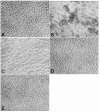
Microscopic evaluation of the erythrophagocytosis induced by polymeric anti-G IgGμtp antibodies. Group O RhD-positive RBC (targets) were sensitised with the different opsonins at a concentration of 3 μg/mL and added to monolayers of thioglycolate-elicited mouse peritoneal macrophages (effectors) at a 5 to 1 target-to-effector ratio. The target and effector cells were examined by light microscopy after incubation at 37 °C for 3 hours. Frame “A” shows a representative image of the control condition (phagocytic cells exposed to unsensitised RBC). The other frames contain representative images taken from samples in which the RBC were sensitised with (B) plasma-derived polyclonal anti-D, (C) monomeric anti-G IgG1, (D) monomeric anti-G IgG3, (E) polymeric anti-G IgG1μtp, or (F) polymeric anti-G IgG3μtp. The images shown (x200) are representative of three independent experiments, in which each condition was tested in triplicate. Ig: immunoglobulin; RBC: red blood cell.
Figure 4
Figure 4
Polymeric anti-G IgGμtp antibodies are better opsonins than plasma-derived polyclonal anti-D. Phagocytic cells and sensitised group O Rh-positive RBC were mixed and incubated as indicated in the legend of Figure 3. After lysing and washing off the non-phagocytosed erythrocytes, the remaining cells (macrophages containing ingested RBC) were lysed and the haemoglobin released was quantified at 630 nm. A standard curve of haemoglobin versus optical density was used to equate absorbance values to RBC numbers, which were converted to a percentage of phagocytosis. Data shown are the arithmetic mean ± standard deviation of three independent experiments, each performed in triplicate. “IgG1μtp=IgG3μtp” means a blend containing equal amounts of each polymeric antibody; “IgG1μtp>IgG3μtp” means a blend containing 75% polymeric IgG1μtp and 25% polymeric IgG3μtp. Statistically significant differences were calculated after Bonferroni’s correction (n= 16) and are indicated with one (p<0.0001) or two asterisks (p<0.001). Ig: immunoglobulin; RBC: red blood cell.
Figure 5
Figure 5
Polymeric anti-G IgGμtp antibodies do not induce complement-mediated haemolysis. Group A RhD-positive (empty bars) or O RhD-positive (black-filled bars) RBC were sensitised with the antibodies indicated in the figure, and then mixed and incubated with autologous plasma as a source of complement. The haemolysis was quantified and normalised to a percentage as detailed in the text and in the online supplementary document. Data shown are the arithmetic mean ± standard deviation of three independent experiments, each performed in triplicate. “IgG1μtp=IgG3μtp” and “IgG1μtp>IgG3μtp” mean the same as indicated in the legend of Figure 4. Statistically significant differences were calculated after Bonferroni’s correction (n= 16) and are indicated with one asterisk (p<0.0001). RBC: red blood cell.
Figure 6
Figure 6
Polymeric anti-G IgGμtp antibodies do not induce C3d deposition on the membrane of RhD-positive RBC. Group A RhD-positive RBC were suspended in HBS-BSA buffer containing (A) no antibody, (B) monoclonal anti-A IgM (a culture supernatant diluted at 1:262,144), (C) monomeric anti-G IgG3 (80 ng/mL), (D) polymeric anti-G IgG1μtp (9 ng/mL), or (E) polymeric anti-G IgG3μtp (9 ng/mL). The sensitised RBC were then incubated with autologous plasma, and finally with an IgM anti-C3d agglutinating reagent. The images shown (X 200) are representative of three independent experiments, in which each condition was tested in triplicate. RBC: red blood cell; HBS-BSA: hepes-buffered saline containing 0.1% bovine serum albumin.

Similar articles

References

    1. Anstee DJ, Tanner MJA. Biochemical aspects of the blood group Rh (Rhesus) antigens. Baillieres Clin Haematol. 1993;6:401–22. - PubMed
    1. Mollison PL, Engelfriet CP, Contreras M. The Rh blood group system (and LW) In: Klein HG, Anstee DJ, editors. Mollison’s Blood Transfusion in Clinical Medicine. 11th ed. Oxford: Blackwell Publishing Ltd; 2005. pp. 163–208.
    1. Kumpel BM. Efficacy of Rh(D) monoclonal antibodies in clinical trials as replacement therapy for prophylactic anti-D immunoglobulin: more questions than answers. Vox Sang. 2007;93:99–111. - PubMed
    1. Bowman J. Rh-immunoglobulin: Rh prophylaxis. Best Pract Res Clin Haematol. 2006;19:27–34. - PubMed
    1. Kent J, Farrel AM, Soothill P. Routine administration of Anti-D: the ethical case for offering pregnant women fetal RHD genotyping and a review of policy and practice. BMC Pregnancy Childbirth. 2014;14:87. - PMC - PubMed

MeSH terms